Medical imaging studies for Tailbone Pain
- Medical imaging studies can be very helpful in making an accurate diagnosis in patients with coccyx pain (also called coccydynia, or tailbone pain).
- The most common medical imaging studies for tailbone pain include:
- X-rays: especially “sitting-versus-standing” x-rays to look for “dynamic instability” ( unstable joints of the coccyx).
- MRI: Magnetic Residents Imaging.
- MRI is helpful at showing soft tissue structures such as pilonidal cysts, retrorectal cysts, abscess (an infected collection of fluid and pus).
- MRI is also helpful when looking for bone infection (osteomyelitis).
- MRI can help in evaluation for possible cancer (malignancy, such as chordoma, which is often fatal and has a tendency to occur at the sacrum and coccyx region).
- CT scans: also called “computerized tomography” scans.
- These can be helpful at showing details of the bone (e.g., the sacrum and coccyx).
- 3-D ( “three-dimensional”) CT scan is a special type of CT scan, where the image can be rotated by the physician, as shown in the video below.
- It is relatively uncommon to need 3-D CT scans in patients with tailbone pain, but in properly chosen patients it can be a VERY helpful imaging test.
Here is Dr. Foye’s VIDEO teaching about 3-D CT scans for Tailbone Pain, Coccyx Pain:
- To reveal the video, click on the image or on the Link below:
- Link to the VIDEO: https://youtu.be/Th6eVSDzfSA
Here is the TEXT from the video:
- OK, I am Dr. Patrick Foye and I’m an M.D., or medical doctor, and I’m Founder and Director of the Coccyx Pain Center or Tailbone Pain Center here in the United States.
- You can find me online at www.TailboneDoctor.com.
- This is just a short educational video on the topic of using CT scan (“cat scan” or computerized tomography scanning) in the evaluation of patients with tailbone pain and specifically the idea of using CT scan with three-dimensional reconstruction, which is what we’re looking at here for this particular individual.
- So the majority of times we are doing our imaging studies for patients with tailbone pain (or coccyx pain, coccydynia) the majority of times we’re using things like x-rays (medically we call those radiographs).
- So standard x-rays look at the tailbone, and especially x-rays done while the patient is sitting versus standing can be hugely helpful.
- In other patients we may need to do advanced imaging studies such as MRI to look at additional detail.
- But about 1% of the patients who I see for tailbone pain may benefit from what’s shown here which is a CT scan (a cat scan, computerized tomography scan) specifically with three-dimensional, or 3D, reconstruction.
- So rather than just having a static image (like one image or one or one slice) what’s done here is actually all of those slices are put together in a digital way, on the computer of course, so that the clinician or a physician can basically rotate the image in multiple directions.
- So I can go back and forth.
- And what that allows me to do is to look from many different angles at a given area, if there’s an area of interest or concern on the patient’s imaging studies.
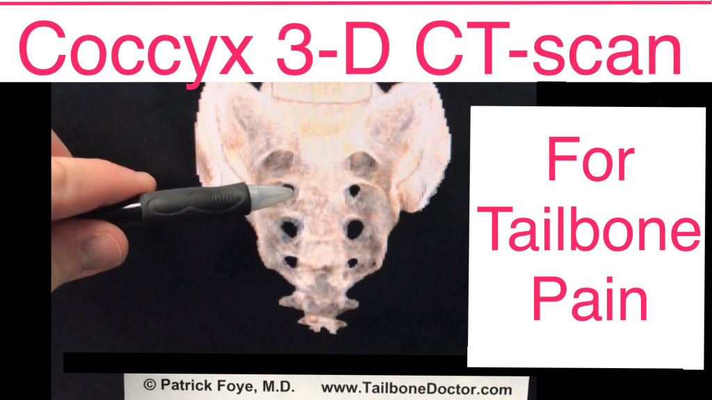
- So just to sort of orient you here on this image…
- So this is the sacrum of this individual up in here, from about here down to about here, that’s the sacrum.
- The sacrum has these holes that you see in it.
- That’s normal.
- These are the sacral foramina which are the holes that the sacral nerves exits through.
- But then we get down to here which is the coccyx or tailbone.
- And one thing that’s particularly interesting here is that in this particular individual you can see that instead of coming to or sort of tapering down to a single point right at the midline…
- we’re going to rotate this and you can see here that this person actually has two different prongs or points that come down rather than just one at the midline.
- So this is somebody who has what’s called a “bifid” (B-I-F-I-D) coccyx or a “bifurcated” coccyx is another name or word for that.
- Really just that describes this anatomical variation where instead of the tailbone coming down to one single point in the midline, instead it splits into a point that comes down on the right and a point that comes down on the left, as shown here.
- This becomes important because if we were looking at this on x-ray…
- well x-ray looking at the x-ray that would be taken face-on (which is called an AP view) a lot of times the AP view of the tailbone is blocked or obscured or made fuzzy because of the structures that are right in front of it blocking the view: so the rectum, the colon, other parts of the organs within the pelvis are there.
- And they can basically (and stool within the within the colon) can make it difficult to see that lower part of the tailbone well on that view on an x-ray.
- But then you go to the side view on the x-ray, which would look something like this (not quite, but similar) and you can see here that because of those two prongs down there, because one is overlapping on top of the other we’re not able to see the space in between them.
- We would have no way to know that there were two separate prongs.
- We would think from this view because they’re overlapping.
- Just like if you were looking at my fingers you’d see two of them here versus now if you really just see one you know what looks like just one finger.
- Whereas being able to rotate brings it out to where you can see the two.
- So the same type of thing here.
- You can see it looks like just one solid lower part of the tailbone.
- But being able to rotate it now we can see that there’s actually two prongs or portions coming down.
- So that becomes really interesting and important because in some patients we see an atomic variations.
- If we’re not sure why we’re not seeing the tailbone appearance in the typical fashion in the lower part of the coccyx, then one of the things that physicians would be concerned about would be whether or not this may represent a malignancy (a cancer) of the tailbone region, such as chordoma, which unfortunately is highly fatal.
- And having a CT scan with 3-D reconstruction like this allows me to rotate the image, bit by bit like this, to look for multiple different angles on the computer screen like this.
- So that, again, I can see from multiple different points of view and decide whether this is just an anatomic variation similar to the way again just rotating my fingers here allows me to see that there’s two fingers rather than just one.
- And the other thing that it allows is that knowing that it’s an anatomic variation then we can move forward with regular treatment of the condition here.
- Or, on the other hand, if this was a malignancy well then we would move forward with getting appropriate further workup or treatment for that.
- So, again, this is maybe one percent of patients who I see for tailbone pain here at the Tailbone Pain Center that I end up ordering this type of study who the CT scan with three-dimensional or 3d reconstruction.
- It’s very cool technology.
- But I only use it in very selected cases who have a specific question anatomically that we were unable to answer from the regular imaging studies (such as x-rays and MRI).
- So that’s all for this.
- This is an educational video, so this is not specific medical advice for any specific individual person or patient.
- For that you would of course seek in-person consultation with your own treating physician.
- If you’re looking for more information on the topic of tailbone pain or coccyx pain certainly you can visit me online at TailboneDoctor.com or on Facebook/TailbonePainCenter.
- So that’s all for now.
- I hope this was helpful.
- If you have questions or comments please feel free to post them down below.
- Bye bye.
For more information on Tailbone Pain, Coccyx Pain: go to www.TailboneDoctor.com
To come to Dr. Foye’s Tailbone Pain Center:
- Get expert medical care for your tailbone problem. Here’s what you need know: https://tailbonedoctor.com/prepare-for-your-visit/
Tailbone Pain Book:
To get your copy of Dr. Foye’s book, “Tailbone Pain Relief Now!” click on this link: www.TailbonePainBook.com

Book: “Tailbone Pain Relief Now! Causes and Treatments for Your Sore or Injured Coccyx” by Patrick Foye, M.D.

Founder and Director at Tailbone Pain Center
Dr. Foye is an expert at treating tailbone pain (coccyx pain).
His personable, private-practice office is located on a modern, renowned, academic medical school campus, at Rutgers New Jersey Medical School.
For an appointment, call 973-972-2802.
https://tailbonedoctor.com/
His personable, private-practice office is located on a modern, renowned, academic medical school campus, at Rutgers New Jersey Medical School.
For an appointment, call 973-972-2802.
https://tailbonedoctor.com/
Latest posts by Patrick Foye, M.D. (see all)
- Coccygectomy: Expected Recovery and Return to Work after surgery for coccyx pain, tailbone pain. - November 28, 2023
- PRP Platelet Rich Plasma or Prolotherapy for Tailbone Pain, Coccyx Pain - October 25, 2023
- Reasons for Normal X-rays and MRI Despite Tailbone Pain, Coccyx Pain - October 3, 2023