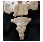
Typical MRI and CT scans are normally done in a horizontal position (while you are lying down).
These tests can provide helpful information, BUT in MANY cases the tailbone will look normal while someone is lying down but will be clearly abnormal/dislocated while the person is sitting with her body weight onto the tailbone.
MRI can be done while sitting, but usually only the “open” MRI facilities can do this and unfortunately those “open” MRI machines typically have a weaker magnets strength which gives a lower quality image.
So x-rays done while sitting are often a better way to go if the goal is to diagnose unstable joints of the coccyx (coccygeal dynamic instability).
- Coccygectomy: Expected Recovery and Return to Work after surgery for coccyx pain, tailbone pain. - November 28, 2023
- PRP Platelet Rich Plasma or Prolotherapy for Tailbone Pain, Coccyx Pain - October 25, 2023
- Reasons for Normal X-rays and MRI Despite Tailbone Pain, Coccyx Pain - October 3, 2023
-Patrick Foye, M.D.
wwww.TailboneDoctor.com


Hi, can you recommend a surgeon with the same specialty in ny/ long island
I had a hemipelvectomy done a year ago, and alot of falls after,my bones are sticking out and extremely painful , my surgeon won’t help me, he was only concerned with the cancer etc… I would love to come there but my insurance is only good in ny, thanks ☺
Hi, Corinne.
First of all, I’m very sorry to hear aboutthe pain and all that you have been going through with the hemi-pelvectomy, etc.
Sorry for the delay in seeing your post.
I do not know of any surgeons in Long Island that perform coccyx surgery.
Definitely feel free to call my office 973-972-2802 and my staff can let you know the details about coming in to be seen. We see many patients from out of state, including those whose insurance plans typically only cover them in their home state.
Hello Dr. Foye,
When there is coccyx pain after an obvious trauma in a female patient, is it reasonable to forgo X-rays? The argument put to me (a female patient who experienced direct trauma to my coccyx) by the ER doctor was that the amount of radiation required to capture a fracture in that location would put reproductive organs at risk. Does that cost-benefit ratio of avoiding X-rays make sense, in this one instance?
I imagine that the same would be true for a CT scan, and if cancer or other causes are unlikely – given a clear trauma – an MRI in the acute stage seems unnecessary, since conservative treatment is recommended initially in any case.
(I am asking because I’d like to know whether it is worth fighting the medical system to get images that might not initially be useful, or might be riskier than they’re worth, in this instance, where the cause is obvious.)
If the ER doctor was right (in my case), does it then make sense to begin with the typical conservative therapy recommended and only pursue imaging and other treatment if it doesn’t work?
Another question – in another post (“Tailbone Fractures take a Long Time to Heal”), you said that “there is no way to avoid putting your body weight on the fracture (unless you totally avoid sitting for several weeks).” If there is in fact an opportunity to totally avoid sitting for several weeks, might it be worth trying? (If so, what kinds of movements and activities would be worth doing to promote recovery?)
A final question – given that in most cases, people continually reinjure or strain their tailbones, can the bones in the coccyx ever *truly* heal? Or is it implicitly understood that the bones will *never* really knit themselves back together, will always be vulnerable, and that the real aim of conservative treatment is pain management? Can manipulation of the tailbone help to “reset” it?
Apologies for so many questions. Your answers would be very much appreciated. The idea of living with this pain for a lifetime – which appears to be quite possible and scarily common – is terrifying.
Hi, Kaylie. While of course I defer to the doctors who are seeing you in-person and who are actually treating you, I am happy to provide general information on the topics that you raise, so you can discuss this with your treating physicians if needed.
You have raised many topics, so I will try to discuss those in order.
1)
Should people forgo initial imaging studies after trauma, in order to avoid radiation to the pelvic/reproductive organs?
This depends.
** For patients with milder trauma or milder pain levels, just having a local bump/bruise/contusion might not warrant any imaging studies at all.
** However, in some cases it is very helpful to know if there is a fracture present. For example, in medical legal cases (such as an injury that happens at work or as a result of an auto accident or falling on the wet floor at a store) your legal case could very much depend upon whether there is objective evidence of injury, and the imaging studies would typically be the best way to document that (and sometimes the only way to document that).
** Also, if a fracture is present then it may change medical management. For example, some medical literature recommends that if there is a fracture the person should initially avoid using NSAIDs (nonsteroidal anti-inflammatory drugs, such as Advil, ibuprofen, etc.). Alternatively, if there is a fracture then it may respond to nasal calcitonin. So it is not really true to say that management is completely unchanged whether there is a fracture or not.
** The x-rays can be done using a coned-down view (putting a collimation extension onto the source of the x-rays). This can dramatically decrease the amount of radiation delivered to the patient, and can especially decrease the amount of radiation delivered to anatomic locations other than the coccyx.
** In terms of radiation exposure, CT scans are typically the worst, giving much more radiation to the patient than x-rays do. MRI does not deliver any radiation to the patient. But MRI is the most expensive so typically it is far more difficult to get an insurance company to pay for an MRI.
2) Meanwhile, yes, in many cases it is reasonably acceptable to start with conservative treatment and if the pain dramatically decreases over the first few days or weeks after the trauma, then perhaps no imaging studies at all will be necessary.
3)
Yes, if you can avoid or minimize sitting on the tailbone that has been injured, then this typically will decrease your symptoms and improve your recovery time.
Meanwhile, although there are activities that make the tailbone pain worse (such as sitting, horseback riding, cycling, motorcycle riding, sit-ups, etc.) there are no specific activities or exercises that help to promote recovery.
4)
Can an injured tailbone “truly heal”?
Yes, injuries at the tailbone can heal just as injuries and bones, ligaments, etc. at other body regions can heal.
Unfortunately, it is also true that in many cases the pain can become persistent and chronic. So medical treatment may be necessary to improve the chance of recovery or at least improve the patient’s pain and quality of life.