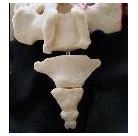
What is a Pilonidal Cyst?
- A pilonidal cyst is a collection of fluid and tissue, which can cause pain in the area of the sacrum and coccyx (tailbone).
- I recently published an online article and video discussing the symptoms and physical exam findings of Pilonidal Cyst.
- You can see that previous article and previous video here: Pilonidal Cyst Causing Tailbone Pain
The next step is to discuss the MRI findings of a Pilonidal Cyst.
- MRI is an advanced imaging test that can show details such as a pilonidal cyst.
- To reveal the video, click on the image or on the Link below:

- Link to the Video: https://youtu.be/QAsZYiKyTyM
Here is the text from the video:
- This is a video just showing a pilonidal cyst on an MRI.
- So basically here, just to orient you, we’re looking at MRI slices.
- We’re looking at the sacroiliac joint on the right and over here on the left.
- And then here’s the sacrum.
- You can just see the start of the where you can see the sacral canal here and a little bit of the sacral hiatus.
- And if I start scrolling down we get down to lower sacrum right where the arrow is here.
- The computer is a little sluggish.
- But coming down here we can see the coccyx on cross-section here at oval-shaped white structure.
- And then here is the pilonidal cyst.
- So this bright white structure on this particular type of MRI image, where basically the white structure here is the pilonidal cyst.
- And then this white line extending to the surface (because out here this is the skin out here, coccyx there).
- So the pilonidal cyst has a “track” or “fistula tract” = the connection or tunnel coming down to the surface of the skin right there.
- Here you can just see the line, that’s the track.
- And the coccyx has now disappeared, so we’re actually now below the coccyx.
- So the pilonidal cyst was right at the back of the coccyx slightly to this side here.
- So this is the midline, so slightly to this side.
- This is actually slightly to the left of midline.
- I know that on the image it looks like it’s to the right, but that’s because the way the patient is laying, the way the image is taken.
- Things over here on this side of the screen will be on the patient’s right side.
- Whereas this side of the screen over here will be the patient’s left side so this is just slightly to the side.
For more information on Coccyx Pain, Tailbone Pain, go to: www.TailboneDoctor.com
To come to Dr. Foye’s Tailbone Pain Center:
- Get expert medical care for your tailbone problem. Here’s what you need know: https://tailbonedoctor.com/prepare-for-your-visit/
Tailbone Pain Book:
To get your copy of Dr. Foye’s book, “Tailbone Pain Relief Now!” click on this link: www.TailbonePainBook.com

Book: “Tailbone Pain Relief Now! Causes and Treatments for Your Sore or Injured Coccyx” by Patrick Foye, M.D.
Latest posts by Patrick Foye, M.D. (see all)
- Coccygectomy: Expected Recovery and Return to Work after surgery for coccyx pain, tailbone pain. - November 28, 2023
- PRP Platelet Rich Plasma or Prolotherapy for Tailbone Pain, Coccyx Pain - October 25, 2023
- Reasons for Normal X-rays and MRI Despite Tailbone Pain, Coccyx Pain - October 3, 2023

