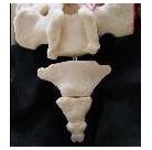
Below are a couple of videos showing how standard x-rays are done at the coccyx (tailbone).
These are sometimes done for patients who have coccyx pain (coccydynia, tailbone pain).
In general, the best x-rays are the sitting-versus-standing x-rays, and you can read more about those here at this link: https://tailbonedoctor.com/sitting-versus-standing-coccyx-x-rays-for-tailbone-pain/
But unfortunately most radiology centers are not familiar with how to do the sitting-versus-standing x-rays. So, in the meantime, patients can at least have the “standard” x-rays done instead. These standard coccyx x-rays typically include lateral views and AP-views, which will be explained below.
The lateral view of the coccyx is typically the most important view when getting x-rays (radiographs) for a patient with tailbone pain.
The “Lateral” view is essentially looking at the coccyx from the side (side view).
Below is a video showing how to do the lateral view xrays of the coccyx:
Below is another video describing how to do the lateral view coccyx x-rays:
And below shows how to do the “AP” view of the coccyx. “AP” stands for Anterior-to-Posterior, which means front-to-back. So, instead of the lateral view (which was the side view) the AP view looks at the coccyx from the front.
Video of the coccyx AP view:
Note: although the demonstration videos show a “patient” wearing denim blue jeans, in reality if this was a real patient we would want her changed into a medical gown. Otherwise, the zippers and denim and grommets or buttons on her pants could block or obstruct or obscure our view of the tailbone.
To get your copy of the book “Tailbone Pain Relief Now!” go to: www.TailboneBook.com or go to Amazon
For more information on coccyx pain, or to be evaluated at Dr. Foye’s Tailbone Pain Center in the United States, go to: www.TailboneDoctor.com

His personable, private-practice office is located on a modern, renowned, academic medical school campus, at Rutgers New Jersey Medical School.
For an appointment, call 973-972-2802.
https://tailbonedoctor.com/