I am honored and excited that I will be giving 3 lectures at the upcoming International Coccyx Pain Symposium in Istanbul, Turkey, July 4-5, 2026.
I encourage pain management physicians, pelvic floor physical therapists, musculoskeletal doctors, surgeons, etc. to join us for a wonderful conference on a topic that is often misunderstood.
(See the video down below if you prefer to watch this as a video.)
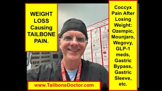
Let’s talk about whether weight loss can actually cause coccyx pain, or tailbone pain.
I’m Dr. Patrick Foye, and I am an M.D. or medical doctor.
I’m the Director of the Coccyx Pain Center or Tailbone Pain Center online at www.TailboneDoctor.com
I’m here in the United States.
One of the questions that comes up sometimes is whether, after weight loss, people can have new onset or worsening of tailbone pain.
This may occur after weight loss surgeries, such as gastric bypass or gastric sleeve.
Or more recently with some of the GLP-1 weight loss medications, such as Ozempic, Mounjaro, or Wegovy.
People will often say, “I lost 50 or 100 pounds or more, and lots of things in my life are much better, but now I have this tailbone pain that I wasn’t having before, or it was never this bad before.”
Let’s talk about a few of the ways that this can happen.
First of all, if we look at this model of the pelvis, here we can see that when somebody sits, they are putting a lot of their body weight onto the sit bones at the bottom of each buttock on the right and left sides.
And not quite as much weight goes onto the tailbone at the midline.
Partly this is because the entire pelvis is elevated somewhat by the gluteal, or buttock, muscles and tissue, including fat within the area.
So people are not sitting quite as directly onto the seat.
But then when someone loses body weight, including losing weight in the gluteal or buttock region, the tailbone becomes that much closer to the chair they are sitting on.
They may never have had tailbone pain before, or it may have only been very very mild or uncommon, infrequent.
But now, all of a sudden, they are having tailbone pain.
Often what is happening can be seen if we look at another model here.
We can see that the tailbone is at the lower end of the spine.
We can see normally there is a small amount of clearance between the tailbone and the chair that you are sitting on, so we are not putting much of our body weight directly onto that area.
But as the pelvis shifts or sinks a little bit lower because someone has lost some of the gluteal body weight, then the lower tip of the tailbone starts to contact the chair, making direct contact in a way that it had not been making before.
So if there is a bone spur there, for example, (which is one of the common sources that I see in patients who travel to see me for tailbone pain after weight loss)… often there may be a bone spur there that can be the cause.
There are special X-rays and MRI studies that can be done in a very particular way to properly image this.
A standard MRI often will fail to show it.
A standard CT scan often will fail to show it.
Even standard X-rays, if they are not done properly with a coned-down collimated view, will fail to show it.
There are particular tests that can be ordered in the proper way, and also carefully on the physical exam, to evaluate for this and other findings that can cause tailbone pain after weight loss.
If you are interested in more information about tailbone pain, then certainly on Amazon you can always grab a copy of my book, Tailbone Pain Relief Now.
If you are interested in coming to see me in person, or find more information and free videos, etc., you can find me online at www.TailboneDoctor.com
Okay, that’s all for now about tailbone pain after weight loss.
In 2008, Patrick Foye, M.D., proposed seated MRI as a new diagnostic test for appropriately selected patients with coccydynia (coccyx pain, tailbone pain).
Publication: Foye PM.A New Diagnostic Test for Coccyx Pain (Tailbone Pain): Seated MRI. American Journal of Physical Medicine and Rehabilitation. 2008 Mar;87(3): S36.
Author: Patrick M. Foye, M.D., Director, Coccyx Pain Center, New Jersey Medical School, 90 Bergen St., DOC-3100, Newark, NJ 07103-2499. Phone: (973)972-2802. Fax: (973)972-2825. www.TailboneDoctor.com
Abstract
BACKGROUND: Diagnostic workup of coccyx pain (tailbone pain, coccydynia) traditionally includes imaging studies such as x-rays, computerized tomography scans, magnetic resonance imaging (MRI), and bone scans. Since coccydynia is often most painful while sitting, a particularly useful technique is to perform lateral radiographs of the coccyx in the seated (weight-bearing) position. These “stress” radiographs may document substantial coccygeal dislocations. However, workup of coccydynia often requires imaging of intrapelvic organs to assess for malignancies and other intrapelvic pathology that may cause referred pain to the coccyx. Meanwhile, increasingly available “positional” or “dynamic” MRI allows MRI to be performed in positions other than traditional supine posture. Thus, the author innovated a new diagnostic test for patients with coccydynia: seated MRI of the pelvis and coccyx. Similar to seated radiographs, seated MRI can document “dynamic instability” of the coccyx, but with the additional advantages of soft tissue imaging of the intrapelvic organs, higher-quality details of coccygeal appearance, and the lack of radiation exposure. This technique has never before been published within the medical literature.
CASE PRESENTATION: The author presents a case of a 37-year old male with coccyx pain after mild trauma. Lumbosacral and pelvic x-rays had been normal. Upon physiatric [rehabilitation physician] pain management consultation after more than one year of persistent coccydynia, we ordered pelvic MRI (to assess for underlying intrapelvic malignancy) and positional (seated versus standing) MRI of the coccyx. Positional MRI documented that while sitting he developed a grade-4 spondylolisthesis between coccygeal segments one and two. His coccygeal dynamic instability on MRI perfectly correlated to his specific symptomatic segment and objectively corroborated his subjective symptoms. Reassuringly, intrapelvic organs were normal.
CONCLUSIONS: The author proposes seated MRI as a new diagnostic test for appropriately selected patients with coccydynia.
(See the video down below if you prefer to watch this as a video.)
This is a short video showing a CT scan, or computerized tomography scan.
A CT scan, or CAT scan, of the pelvis, particularly focusing on the sacrum and coccyx, in a patient who underwent a coccygectomy. And the first coccygeal bone (which is this bone right here) was essentially left behind.
So it’s a “partial” coccygectomy, rather than a complete coccygectomy.
I’m Dr. Patrick Foye. I’m the Director of the Coccyx Pain Center, online at www.TailboneDoctor.com.
In this short video, we’re going to review this.
The reason why we know that this is indeed the first coccygeal bone is from looking at the anatomy.
In my book, I have a whole chapter on anatomy.
I’m holding the camera with one hand, so I’m trying to show you with the other hand.
In the chapter on anatomy, if you look here, you can see that the first coccygeal bone has these two areas.
These two areas come up—they call them horns or cornua—that extend upward from the top of the coccyx.
Those meet the sacral cornua, which project downward.
From the side view, we can see that the cornua hook up like that.
Coming back to our CT scan here, you can see those cornua right here, coming upwards from that remaining coccygeal bone.
If I swing around, you can see them very nicely here again—this one and this one—projecting upwards.
Those are signs that this is indeed the first coccygeal bone.
The coccygeal cornua, or horns, are projecting upward to meet the sacral cornua, which are projecting downward.
This is where the first coccygeal bone was left behind.
I’ll show you that in one other spot here on an anatomic model of the coccyx.
Again, you can see those horns or cornua heading upward, just as we see here.
That is indeed the first coccygeal bone still remaining in this patient who underwent a coccygectomy.
Unfortunately, there are many times where patients are told that they are having a complete coccygectomy, but sometimes the first coccygeal bone or other coccygeal bones may be accidentally or inadvertently left behind.
Then, we need to decide whether they should have been removed.
Most people do not need coccygectomy surgery for their tailbone pain.
But for those who do, it needs to be a conscious decision made between the patient and the surgeon about whether to do a total versus a partial coccygectomy.
If the decision is to do a total coccygectomy, then you want to make sure that the entire tailbone is removed and that a piece of it is not accidentally left behind.
I just wanted to point that out since this is a good image for showing that.
That’s all for now.
I hope this has been helpful in distinguishing partial versus complete coccygectomy.
For more information, you can find lots of details in my book, Tailbone Pain Relief Now, which you can get online at Amazon.
I hope this info is helpful for you.
If you need more information about tailbone pain, you can find me online at www.TailboneDoctor.com.
Using fluoroscopic guidance (fluoroscopy) helps doctors when doing a coccyx injection (tailbone injection) treat coccyx pain (tailbone pain, coccydynia).
(See the video down below if you prefer to watch this as a video.)
This video is about the use of fluoroscopy, or fluoroscopic guidance, when performing coccyx injections or tailbone injections.
I’m Dr. Patrick Foye. I’m an M.D., or medical doctor, and I’m the Director of the Coccyx Pain Center, or Tailbone Pain Center, here in the United States. My website online is www.TailboneDoctor.com.
In this short video, we’re just going to talk a little bit about the benefits of using fluoroscopic guidance when doing or having coccyx injections in the treatment of coccyx pain or tailbone pain.
Behind me here is the fluoroscopy machine, as shown here, with the screens up there.
Fluoroscopy is similar in some ways to X-ray, which can be displayed up on a computer screen, except it’s done in very tiny increments—often just a fraction of a second per image.
When a patient is having an injection for coccyx pain, the very first thing—more important than the injection itself—is that the physician actually does his or her best to make an accurate diagnosis.
I see this all the time in patients who fly in to see me from around the country.
They’ve had multiple injections done for tailbone pain, but nobody has actually made a diagnosis as to what’s causing their tailbone pain.
That’s a problem because, if you don’t have a specific diagnosis, it can be more difficult for the physician to know the target location at or around the tailbone where the injection should be done.
Assuming that you’ve gone through the appropriate steps to have a specific diagnosis made (this typically includes having coccyx X-rays done while you are sitting and comparing those with the position of the coccyx or tailbone while you are standing)…
Let’s assume that you’ve had those sitting and standing X-rays done, and they showed a dislocating segment between coccyx bone number three and coccyx bone number four.
In that case, you would want the injection to target that specific joint, bone, or location where the abnormality was found.
Unfortunately, what I often see is that physicians will either do the injection “blind,” meaning they’re not using fluoroscopic guidance—they’re just sticking the needle somewhere in the area of the coccyx and injecting steroid, lidocaine, or another local anesthetic, and just hoping for the best.
Sometimes, that may actually give some benefit for some patients, but it is far from ideal.
Ideally, you want to have a specific diagnosis when possible (which it is in the majority of patients), and then you want to target the injection to the specific site of abnormality.
Using the fluoroscopy machine, we can target the specific joint, bone, or abnormality.
Just to show you a little bit of how this works, this is the fluoroscopy machine right here, with the monitors up here behind me.
On the table here, I have a plastic model of a pelvis.
You can see that the coccyx is right down here at the lower part of the spine.
When I take the fluoroscopic image, as shown here, you can see the details of the coccyx.
In this case, it’s a plastic model of a coccyx, but it still shows the individual bony segments and the joint spaces between them.
Here’s the lower tip down here.
For many patients, there will be a bone spur at the lower tip of the tailbone.
That diagnosis needs to be made from the patient’s X-rays, MRI, or CT scan, or sometimes it can even be detected on physical examination.
Then, you want the injection to be specific to the patient’s abnormality.
By seeing the details on the fluoroscopy screen, I can ensure that I place the needle precisely at the joint, bone, or abnormality in question.
This way, I can be confident that I’ve achieved good coverage in the correct area.
Unfortunately, many patients have injections done by physicians who are not as familiar with treating the tailbone.
They may either perform the injection blindly or, even if they use fluoroscopy, they haven’t properly identified the abnormality.
As a result, they use fluoroscopy to inject somewhere on the coccyx, but often it’s not the exact place where the patient actually has the abnormality or truly needs the injection.
This just gives you a general idea of the setup for an injection.
The patient comes in, we have a fresh sheet on the table, we position them appropriately, and we get the fluoroscopic image displayed on the computer screen.
Then, we perform the injection accordingly.
That’s just a little bit about fluoroscopic guidance when it comes to performing coccyx injections.
If you’re interested in more information about coccyx pain, you can visit my website at www.TailboneDoctor.com.
If you have tailbone pain and are interested in getting a copy of my book, “Tailbone Pain Relief Now”, you can find it at www.TailboneBook.com, or you can search for “Tailbone Pain Relief Now” on Amazon.
If you search for “tailbone” along with my last name, “Foye,” the book will come up in the Amazon search results.
If you’re interested in coming to see me for evaluation and treatment for tailbone pain, the easiest way to find me is through my website, www.TailboneDoctor.com.
I hope this has been helpful information about the importance and benefits of using fluoroscopic guidance for injections for tailbone pain.
Okay, bye-bye.
Here is the video:
Here is a photo screenshot from the video:
Fluoroscopic Guidance for Coccyx Injections for Tailbone Pain, coccyx Pain, Dr Foye
To learn more about image-guidance for tailbone injections, go to this Link:
Rowing machines are a fantastic way to get a full-body workout, but they can also be the culprits behind coccyx pain (tailbone pain). Understanding how this happens and what you can do to prevent it can help you enjoy your workout without discomfort.
What Causes Coccyx Pain from Rowing?
Prolonged Pressure: The most common cause of coccyx pain is prolonged pressure on the tailbone. When you’re rowing, you’re sitting on a hard surface for an extended period, which can lead to discomfort and pain.
Repetitive Motion: The repetitive nature of rowing can also contribute to coccyx pain. Continuous rocking back and forth on the coccyx can cause or worsen tailbone pain.
Poor Posture: If you’re not sitting correctly or maintaining proper form, you might be putting additional stress on your coccyx. Slouching or leaning too far back can exacerbate the problem.
Inadequate Padding: Many rowing machines come with hard seats that don’t provide enough cushioning. Without proper padding, the tailbone is more susceptible to pain.
Symptoms to Watch For
Discomfort When Sitting: The most common symptom is pain at the lowest tip of your spine while sitting. Pain may increase while you are sitting, especially while leaning partly backwards, or during the first few moments when you go from sitting to standing.
Tenderness to the touch: It may be painful when you press on the coccyx. The coccyx (tailbone) is the bony area just above the anus. The pain can range from a dull ache to sharp, stabbing pain.
Radiating Pain: The pain might also radiate to your buttocks and pelvis.
Tips to Prevent Coccyx Pain
Adjust Your Position: Make sure you’re sitting properly on the rowing machine. Sit upright, engage your core, and avoid slouching.
Use Padding: Consider using a cushioned seat cover or a padded rowing machine seat to reduce pressure on your coccyx. You may want to use a coccyx cushion on top of the seat, to minimize pressure on the tailbone.
Take Breaks: Don’t stay seated for too long. Take regular breaks to stand up, stretch, and relieve pressure.
What to Do if You Experience Coccyx Pain
Rest: If you start to feel pain, take a break from rowing. Resting can help reduce inflammation and give your body time to heal.
Apply Ice: Ice can help reduce swelling and numb the area, providing relief from pain.
Over-the-Counter Pain Relief: Non-prescription pain relievers like ibuprofen may help manage discomfort. But oral medications can have side-effects, so be careful not to take them too frequently.
Seek Medical Advice: If the pain persists or is severe, it’s a good idea to consult with a healthcare professional. They can offer more specific advice and treatment options. It is especially important to find a doctor with expertise specifically in evaluating and treating tailbone pain, since most doctors do not have much experience with this condition. An experienced physician will know if certain types of pain management injections may be helpful.
Consider alternative exercises: If tailbone pain prevents you from rowing, consider other types of exercises that will allow you to stay healthy without causing pressure and pain at the coccyx.
Conclusion
Rowing is a great way to stay fit, but it’s important to be mindful of your coccyx. By understanding the causes of tailbone pain and taking steps to prevent it, you can enjoy your workouts without discomfort. Always listen to your body and make adjustments as needed to ensure a comfortable and safe exercise routine. Happy rowing!
Raise awareness of tailbone pain (coccyx pain) and advocate for patients suffering from this.
Provide educational resources to support people with tailbone pain, helping them find useful information about the most modern tests and treatments.
Educate healthcare providers, as tailbone pain is often under-diagnosed and poorly understood, since most doctors have little or no training about this condition.
History:
Established in 2018 by Patrick Foye, M.D., the Founder and Director of the Tailbone Pain Center at Rutgers New Jersey Medical School.
Dr. Foye created this day after years of treating thousands of patients with tailbone pain, aiming to bring attention to a condition that is frequently overlooked and misunderstood.
Events planned for November 13, 2024:
Free book: all day on Amazon, you can get a free copy of Dr. Foye’s book “Tailbone Pain Relief Now! Causes and Treatments for Your Sore or Injured Coccyx.” The e-book version free on via Amazon, worldwide.
Free Facebook Live question and answer session all about tailbone pain. Dr. Foye will host a live session on www.facebook.com/TailbonePainCenter/ to answer questions coccyx pain, including symptoms, diagnostic tests, treatments, etc. That live session will be: Wednesday, November 13, 2024, at 7pm eastern time zone (New York City time zone). You can post your questions in advance, or post them live in the chat during the event. Here is the link: https://www.facebook.com/share/15Z1h1sEpc/
Before mobilizing (manipulating) the coccyx, FIRST the treating physicians should DIAGNOSE what is CAUSING the tailbone pain.
The most common cause of coccyx pain is coccygeal HYPERMOBILITY. Hypermobility means that there is too much mobility, or movement, at the bones and joints of the coccyx. This is caused by laxity, or looseness of the ligaments that would normally keep the joints stable. This is also called coccygeal dynamic instability. The important point is that this is the MOST COMMON cause of coccyx pain.
Be careful, since if the coccyx ALREADY has excessive mobility, then mobilization (manipulation) could make the hypermobility even WORSE.
If the coccygeal joints are already too loose, moving too much, then it probably does not make sense to do a treatment to make the joints move even more or to loosen those joints even more. Manipulation can make the hypermobility even worse.
To diagnose hypermobility: Sitting-versus-standing x-rays of the coccyx are done to assess whether there is excessive movement of the coccyx while the person is sitting. When sitting, you put your body weight onto the coccyx. The tailbone position while sitting is compared with the position while standing. For more information on this, see: https://tailbonedoctor.com/sitting-versus-standing-coccyx-x-rays-for-tailbone-pain/
The important point is that the treating physician should work to make a specific diagnosis FIRST, BEFORE considering manipulation / mobilization of the coccyx.
COME FOR RELIEF: For more information on coccyx pain, or to be evaluated in-person at Dr. Foye’s Coccyx Pain Center in the United States, go to: www.TailboneDoctor.com
– Patrick Foye, M.D., Director of the Tailbone Pain Center, New Jersey, United States.
(See the video below if you prefer to watch this as a video.)
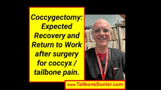
Let’s talk about what to expect for recovery after coccyx removal surgery which is called coccygectomy.
I’m Dr. Foye, the director of the Coccyx Pain Center or Tailbone Pain Center here in the United States at Rutgers New Jersey Medical School and I’ve been treating patients with coccyx pain for about 25 years, thousands of patients.
The good news is that the vast majority of patients do not require surgical amputation for treatment of tailbone pain.
Most people do respond well to non-surgical treatment.
However, there is a small percentage of patients who may require surgery.
And for those patients who do require surgery (or that I send for a surgical consult) they always ask what should I expect after surgery? what’s the recovery like?
So let’s just talk a couple of minutes about that.
It really varies of course from patient to patient.
And that’s dependent on a couple of important factors.
Number one is the skin and how well it heals at the surgical site.
For some patients the skin heals nicely smoothly without any difficulty.
Other patients unfortunately run into problems with skin breakdown at the surgical site, where it may get infected or the skin just may not heal well. or the scar may open up partly because it’s an area that we sit on.
So there’s a lot of restrictions in terms of how many weeks or months until you can sit on the tailbone area after you’ve had the coccyx or tailbone surgically amputated or removed.
Another issue then is the pain at the area.
Some patients are able to tolerate sitting earlier than others.
So for some people it’s hey I’m only a couple of weeks out from surgery and I can already tolerate sitting for short periods of time, such as 10 or 15 minutes.
For other patients it may be months and months out and they’re still having a lot of pain or discomfort while sitting. This can be either because of scar tissue or because of a retained bone fragment or just the skin not healing well or those kinds of things.
So again, it runs the spectrum in terms of outcomes.
Other factors are things like what kind of job is the person trying to get back to.
So for example some jobs require sitting a lot of the time, without a lot of ability to get up and stand during the work day.
So if you think about a job like an airline pilot or a bus driver where once they sit in that chair they need to be sitting in the chair for the bulk of the time that they’re doing their job.
They’re not able to just stand up at will throughout the workday like people in other occupations might be able to.
Another factor is how long is the commute to work.
If we’re talking about return to work expectations, if somebody is working from home and they’re able to do to stand up while they do a video meeting or something like that, then obviously the chances of them getting back to work soon is much better than for somebody who has an hour and a half drive for their commute each way and thus they may have a really difficult time tolerating the drive to and from work, let alone the sitting that they may need to do when they’re actually at work.
In general, it’s going to be important for you to work closely with the surgeon and their surgical office or team.
I’m not a surgeon, so I most I spend most of my time and professional career helping people avoid going for surgery when they can.
But if you do go for surgery, it’s important to work with your surgical team.
Make sure it’s clear to them if you’re having any difficulties or challenges.
Ask them what their specific advice is for how soon you should be able to sit, how long you should be able to sit, when you should be able to go back to work, and those kinds of things.
The biggest advice I could give you is to make sure that number one of course work with your surgeon.
But number two that you go gradually.
The biggest mistake I see people make is that they’re happy that they finally had the tailbone removed (if they’re getting a good initial outcome) but then they either go back to work too soon or they start doing prolonged sitting.
Or they think well the tailbone is gone maybe I can sit on an exercise bike and try it for 20 minutes.
And really my concern there is that it’s very easy to overdo it and have a big setback.
I would rather see people go more gradually and have a nice smooth recovery where they can sit for longer and longer periods of time.
So hopefully this video is helpful just for helping you to have some general expectations for things to take into consideration if you are having tailbone removal surgery (coccygectomy).
Now you have some idea about the variability and the factors that go into what recovery time frame is like and how long until you can sit again (we’re talking usually 6 weeks 8 weeks and then it’s going to be even a gradual progression after that).
And for return to work again that’s going to be very depending on the job and the commute and those kinds of things.
And coccygectomy is really known as a surgery that has a much longer recovery time than most surgeries.
So, it’s not the type of thing where hey I had this the tailbone removed that was yesterday today I’m totally fine and going about my full life’s activities.
That would be naive to go in thinking that’s going to be the outcome.
So hopefully this gives you some context for that.
Some people say it takes 6 to 12 months before fully assessing response to coccygectomy.
If you want more information you can find me online at www.TailboneDoctor.com you can grab a copy of my book on Amazon.
COME FOR RELIEF: For more information on coccyx pain, or to be evaluated in-person by Dr. Foye’s Coccyx Pain Center in the United States, go to: www.TailboneDoctor.com
– Patrick Foye, M.D., Director of the Tailbone Pain Center, New Jersey, United States.
Below, is the screen-capture image from the video: