When: Wednesday, May 9, 2018, at 12:00 noon eastern time (New York City time).
Why: I already am on Twitter, Google+, Facebook, Pinterest. Reddit is a way to educate a new group of people about tailbone pain.
Limitations: I generally avoid giving actual individual medical advice online to those who I have not evaluated in person. But I can provide information and answers that you can discuss with your in-person treating physician.
Watch the VIDEO:
Here is the screenshot thumbnail image for the video:
Reddit, AMA Ask Me Anything about Tailbone Pain, Coccyx Pain, Coccydynia
And this is the next in this series of videos going chapter by chapter through my book Tailbone Pain Relief Now.
The idea is to give you highlights of each chapter and provide a format where we can interact and have questions and conversations about the chapter by putting your comments down below the video.
So here now we are up to chapter number 12, which is Sympathetic Nervous System Pain at the Coccyx ,or causing tailbone pain essentially.
And the idea here is that our pain pathways are set up to sort of sound an alarm system.
So if I touch my hand to a hot stove, the pain will signal me to pull my hand away from the stove and then the pain should stop.
But sometimes at the tailbone what happens is that somebody has a bone spur or a dislocating bony segment or arthritis and the pain doesn’t stop.
The pain is painful every time the person sits.
They’re not able to get relief either because their local doctors are not able to give them an accurate diagnosis or an effective treatment plan.
So the pain goes on and on day after day week after week month after month.
And after a while, the nerves themselves can become hypersensitive and hyper irritable.
And at the tailbone there’s a particular type of nerve structure that’s there that’s part of what’s called the “sympathetic nervous system.”
And the “sympathetic” nervous system has nothing to do with feeling “sympathy” or empathy for the person that’s having pain.
It’s just the medical term for that type of nerve pathway.
The sympathetic nervous system is known as the part of the fight-or-flight response.
So the idea is that if for example thousands of years ago if a saber-toothed tiger or something were to attack us as humans, then we would have a response where we’re either going to fight it off or we’re going to run away. So, fight or flight.
And the idea is that when we have a perceived threat we’re going to react to that.
And lots of things happen as part of that sympathetic nervous system: our blood pressure goes up, our heart rate goes up as our heart beats faster to get more blood out to our brain and to our muscles and our pupils get bigger so we can look around and assess the threats.
All kinds of chemicals are released in our body. So things like adrenaline, epinephrine is sort of running through our system.
So it’s really this sympathetic nervous system response that happens.
Now, very interestingly, the entire sympathetic nervous system has this pathway where the sympathetic nervous system is right along the spine on each side right and left.
But when it gets down to the tailbone instead of having a right and left ganglion or hub for each of the stopping points along the way for the sympathetic nervous system, there’s just one at the midline and that’s called the ganglion impar.
And impar means solitary or unpaired because it does not have a right and a left.
It has just the one at the midline and that’s located right at the level of the upper coccyx.
So now you have this pathway for sort of sounding the alarm when there’s a threat to you.
And the very final train stop (if we want to call the ganglion that)… the very final hub or stop along the way of the sympathetic nervous system ganglion (those chains of nerves that are linked together)… the very final one is right at the front of the tailbone.
So you can imagine that if there’s a cause of tailbone pain such as a dislocation, an unstable joint, arthritis in the joint etc, that the local pain driving that irritation in the area can start to have a phenomenon where in addition to the musculoskeletal cause of pain, there can actually also be a nerve pain on top of that.
So nerves are hyperirritability or hypersensitivity in that area.
So that’s part of the sympathetic nervous system that can be painful at the coccyx.
And this becomes really important because if you only treat the musculoskeletal cause of pain without also treating the nerve pain then to the patient they just know that they still have pain.
And the doctor and the patient maybe are not aware of why the pain is persisting.
So, maybe there was a bone spur or arthritis or a dislocating segment and perhaps there was an anti-inflammatory injection done to help with the musculoskeletal pain and inflammation.
But the pain persists and the pain persists in those cases perhaps because the sympathetic nervous system is irritated and nothing was done to quiet that down as well.
So, often it can be helpful to, in addition to treating the musculoskeletal cause of the pain, to also put some local anaesthetic such as Lidocaine, etc., on that sympathetic nerve ganglion, that ganglion impar at the coccyx.
How you would do that depends. It needs to be “custom done” essentially depending on the specific anatomy of a given patient.
I’ve published a number of different techniques for doing this.
The original publication was way back in the 1990s by Dr Plancart down in Mexico City.
But I’ve more recently published other techniques.
This is an area I lecture on quite a bit.
But basically, that’s the idea as far as the ganglion impar and sympathetic nervous system pain.
Other examples of sympathetic nervous system pain that happen in the body… sometimes people may be familiar with things like R.S.D. or Reflex Sympathetic Dystrophy or Complex Regional Pain Syndrome (sometimes abbreviated as C.R.P.S.).
Those are conditions where there’s essentially a hyperactive, irritable sympathetic nervous system causing pain usually in an arm or a leg.
And doing a local anesthetic block for that limb, that arm or leg, can give a lot of relief.
And, similarly, doing a sympathetic block for patients with a sympathetic nervous system pain at the tailbone can give a lot of relief in that area.
So that’s the general idea.
There is a lot more information, of course, within the chapter, within the book.
So, again, Chapter 12 about that type of nerve pain at the coccyx.
If you have further questions or comments on that, definitely post them in the comments down below.
I’ll be interested to read those and respond to those and I’m sure others will find your comments helpful as well.
If you are looking for a copy of the book, the easiest way to get that is to go online at www.TailboneBook.com
And from that web page I have the links to the Amazon sites in different countries and such that you would use to purchase the book, depending on where you are located.
You can get the paperback book, the whole thing is two hundred and seventy-two pages.
Or you can get it as an e-book, an electronic book which you can basically download for a couple of dollars. And that’s available anywhere in the world where you have internet access. You can get the electronic book and you can read that online, you don’t need any special device other than however you access the internet.
So anyway, I hope that that information is helpful for you.
If you have questions, again, post them down below.
To get the book, go to www.TailboneBook.com.
And to find me online or to come for an evaluation here at the Coccyx Pain Center, you can find me by going to www.TailboneDoctor.com.
All right, I hope that’s helpful.
Bye-bye now.
Here is the actual VIDEO:
Here is the screenshot thumbnail image for the video:
Chapter 12 of Tailbone Pain Book, Sympathetic Nervous System Pain of the Coccyx, Causing Tailbone Pain, Coccyx Pain
To get your copy of the book “Tailbone Pain Relief Now!” go to: www.TailboneBook.com
For more information on coccyx pain, or to be evaluated at Dr. Foye’s Tailbone Pain Center in the United States, go to: www.TailboneDoctor.com
Below (at the bottom of this page) is a VIDEO of Dr. Patrick Foye, M.D., who will be giving lectures at the conference.
Dr. Foye will be giving two lectures on non-surgical treatments for coccyx pain (tailbone pain, coccydynia).
Dr. Foye will post updated information from the conference, either during or after the coccyx, as much as possible.
Here is the text from Dr. Foye’s video:
This video is about the upcoming second International Coccyx Pain Symposium, which will be taking place in the end of June 2018 in the Netherlands.
I’m Dr. Patrick Foye. I’m an M.D. or Medical Doctor and Director of the Coccyx Pain Center here in the United States.
I’m online at www.TailboneDoctor.com.
And this upcoming conference is online at www.Coccyx Symposium2018.com.
So, the first International Coccyx Pain Symposium was two years ago in 2016, that was in Paris, France.
It was a terrific success. I got to meet clinicians and researchers, anatomy folks etc, from literally around the world who came and spoke at or attended the first International Coccyx Pain Symposium, in Paris.
And that was really, really terrific.
People whose research I have been reading for years… to be able to meet them in person and really learn a lot from different people in different specialties.
And I’m really optimistic that this second International Coccyx Pain Symposium will be a terrific success as well.
There is a wide variety of different speakers represented, both physicians and non-physicians and within physicians there are surgeons and non surgeons and other specialties.
So it really gives a diverse bunch of different viewpoints as to how different doctors or clinicians in different specialties may approach patients who have similar problems.
And really that’s how we all learn from each other.
Coccyx pain is a rare and uncommon enough condition that for many of us who treat this we don’t have a lot of other physicians around us who have a lot of experience in treating this as well.
So it’s really valuable to have a conference like this where many of us from around the world get together and exchange ideas about the best ways that we can help these patients.
The speakers: there’s more than a dozen speakers from literally around the world.
So right from within the Netherlands there are a number of physical therapists who be speaking so Pelvic Floor Physical Therapist for example.
There is a Colorectal Surgeon from the Netherlands as well.
There are Orthopedic Surgeons from both Paris, France, and also elsewhere in France, and two Orthopedic Surgeons will be coming down from Norway.
There is a Physical and Manual Medicine and Rheumatology Physician from Paris, France, who is certainly well known for his work in coccyx pain, he will be speaking as well.
There is a Physical Medicine Rehabilitation and Pain Physician from Turkey who will be speaking, an Anesthesiology Pain Physician from the United Kingdom who will be speaking, a Chiropractor from the UK as well, a Physicist from the UK who is a patient advocate representing the patient perspective for people suffering with tailbone pain, there is an anatomy PhD researcher who is coming all the way from New Zealand who specializes in some of his research specifically about issues related to the anatomy of the tailbone.
And I’m sure there’s others I’m forgetting as well.
I’ll be speaking, giving a couple of lectures as well,.
So I’ll be coming from the United States.
So again, a wide variety of speakers.
I’m really looking forward to the conference and I’ll be of course posting information from the conference and maybe doing some live streaming if I have adequate Wi-Fi while I’m there to do so.
So if you’re interested in the conference, it’s mostly attended by clinicians but if you have an interest in the coccyx or tailbone pain in general you may be interested in attending, if you’re anywhere able to reach the Netherlands in the end of June.
Again, the information for that is online at www.CoccyxSymposium2018.com.
Or I’ll be putting more information up about it as it’s upcoming and as I attend the conference myself.
Should the Coccyx Bones be Fused Together? Does that Cause Tailbone Pain?
There is a lot of variability from person to person regarding fusion of the coccygeal joints.
The Completely Fused Coccyx: A Rigid, Non-mobile Tailbone
In some people, the entire coccyx is fused, meaning that the individual bones of the coccyx are fused into one solid bone.
When this happens, there is no mobility or movement possible. In that situation, the tailbone is stiff or rigid.
A stiff, rigid coccyx is not able to move out of the way when you sit. So, it may be more prone to pressure and pain while sitting.
The Completely Patent Coccyx: a Tailbone where None of the Joints are Fused
In other people, the individual joints may all be patent (not fused).
When those joints are patent, it means that there can be movement at those joints.
The movement of those joints could be either normal or abnormal, depending on how sturdy the ligaments are.
Ligaments attach one bone to the next bone. If the ligaments are torn, stretched, or loose, then there can be excessive movement at the joint (also called hyper mobility or instability).
The best way to assess for hypermobility is by doing sitting versus standing x-rays and comparing the position of the tailbone while someone is standing compared with while they are sitting. You can read more about sit-stand coccyx x-rays here at this link.
A Coccyx where SOME Joints are Fused and SOME Joints are Patent
Many people have some of their coccygeal bones fused together, while some of their other joints at the coccyx are not fused.
The take home message is that this varies from patient to patient.
It is important to CORRELATE the imaging findings with the exact site of the patient’s pain.
To get your copy of the book “Tailbone Pain Relief Now!” go to: www.TailboneBook.com
For more information on coccyx pain, or to be evaluated at Dr. Foye’s Tailbone Pain Center in the United States, go to: www.TailboneDoctor.com
Fortunately, someone with tailbone pain in the UK looked into this further and shared the following information with me. They do not want to be named or given public credit, so I will of course respect their privacy. But I do acknowledge and greatly appreciate their efforts and the information they provided, which is below.
They tracked down the Royal College of Radiologists (RCR) latest national guidelines, called iRefer. “They list conditions and recommended imaging, with comments. These form the basis of Trust guidelines.”
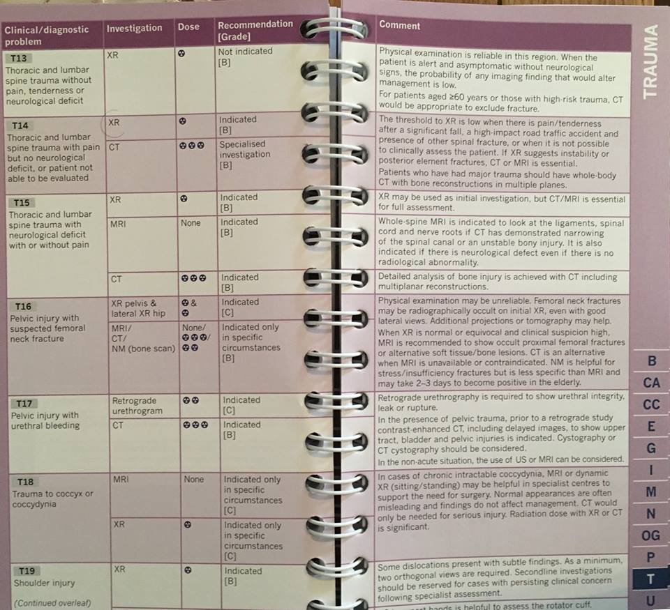
“So I spent £30 on the latest set of iRefer RCR guidelines…. Lo and behold, as of December 2017, they do recommend sit/stand x-rays in intractable cases (SEE PHOTO). They mention that these need to be done at specialist centres. We need to find out where these centres are – ask your local spinal surgeon about this. Your GP [general practicioner] is unlikely to know as it is a specialist investigation. I suspect that the message has not trickled down to all radiology departments… and guidelines at most Trusts haven’t been updated yet.”
Summary on UK coccyx x-rays:
Good News: In the UK, the Royal College of Radiologists does have guidelines (from year 2017) that do indeed recommend sit/stand x-rays of the coccyx in intractable cases of coccyx pain.
Bad News: In the UK (and much of the world), you still may have a challenging time finding a radiology center that knows how to properly perform and interpret the sitting-versus-standing xrays.
Those in the UK suffering from coccyx pain should share this Royal College of Radiologists iRefer guidelines with their treating doctors.
Photos of the Royal College of Radiologists iRefer guidelines book, section on coccyx x-rays for tailbone pain:
Royal College of Radiologists, RCR, iRefer, Guidelines
Sit-Stand-Coccyx-Xrays, supported by UK criteria from the Royal College of Radiologists, RCR, iRefer, Guidelines
Sit-Stand-Coccyx-Xrays, supported via UK criteria from the Royal College of Radiologists, RCR, iRefer, Guidelines
To get your copy of the book “Tailbone Pain Relief Now!” go to: www.TailboneBook.com
For more information on coccyx pain, or to be evaluated at Dr. Foye’s Tailbone Pain Center in the United States, go to: www.TailboneDoctor.com
Here’s an idea to help improve surgical outcomes when coccygectomy is done for coccydynia, coccyx pain, tailbone pain:
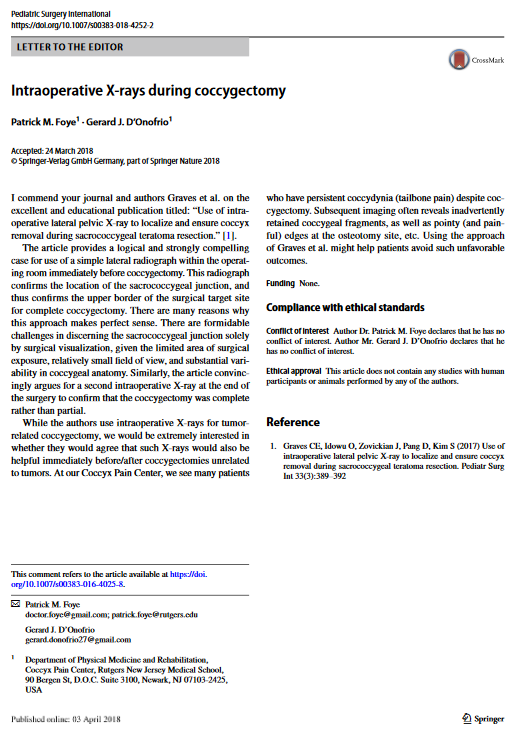
Intraoperative X-rays During Coccygectomy, To Improve Surgical Outcomes for Coccydynia, Coccyx Pain, Tailbone Pain
I wrote this up and submitted it to a medical journal called “Pediatric Surgery International” and they published it this past week.
Over the past 20 years, many patients have come to see me due to persistent coccyx pain after coccygectomy (surgical amputation of the coccyx).
In many cases, we find that the surgeon accidentally left fragments of the coccyx behind. Depending on the location, these can be painful.
In other cases, the primary site of the coccyx problem was never removed by the surgery.So the pain persists.
These problems happen mainly because the surgeon has only a limited view through a small incision during this surgery into a small area.
My proposed solution is for surgeons to take x-rays immediately before starting the surgery and before ending the surgery. This way, the surgeons could be more confident about whether they removed all of the bones that they wanted to remove. The x-rays would also show if the surgeons left a sharp or pointy edge at the site where they cut through the bone. (This is important since a sharp, pointy edge can cause pain when you sit on it.)
I brought this up in the context of a case where x-rays were used during removal of a cancer in the coccyx region. My proposal is that this approach should also be used for coccydynia patients who do not have cancer.
I am very happy that this international surgery journal has published this.
I hope that surgeons will read this and start to follow this recommendation.
I think this can decrease the number of coccygectomy patients who have a bad and painful outcome.
Dr Foye’s Letter- X-rays during coccygectomy for coccyx pain, tailbone pain
PubMed – X-rays during coccygectomy for coccyx pain, tailbone pain
Pediatric Surgery International, Journal Cover
Intraoperative X-rays during coccygectomy, Regarding Coccyx Pain, Tailbone Pain
To get your copy of the book “Tailbone Pain Relief Now!” go to: www.TailboneBook.com
For more information on coccyx pain, or to be evaluated at Dr. Foye’s Tailbone Pain Center in the United States, go to: www.TailboneDoctor.com
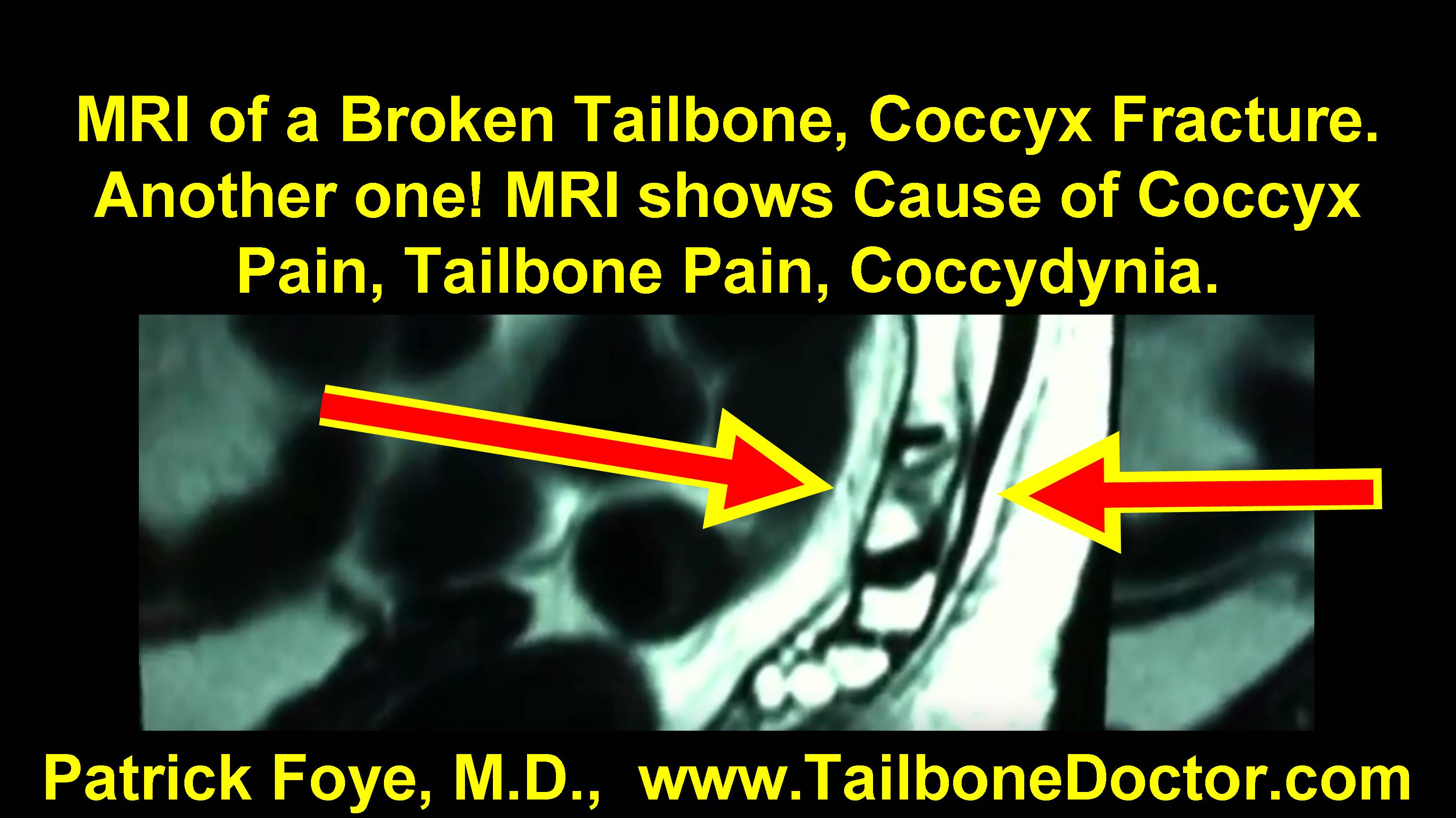
MRI Tips on How To See a Broken Tailbone, Coccyx Fracture.
You Can See what the Radiologist Missed!
Patrick Foye, M.D. discusses MRI (Magnetic Resonance Imaging) showing of a Broken Coccyx, or Tailbone Fracture.
MRI shows a Fractured Coccyx.
Radiologists often FAIL to see these abnormalities!
The actual VIDEO is at the bottom of this page.
Here is the TEXT from the video:
Okay. This is just a short video showing a fracture of the tailbone, a coccyx fracture or tailbone fracture or a broken tailbone, broken coccyx, however you want to say it.
This is a coccyx injury that has resulted in a bony fracture at the tailbone.
I am Dr. Patrick Foye.
I am Director of the Coccyx Pain Center here in New Jersey.
Just a quick F.Y.I., these videos are meant of course purely for educational purposes, they are not to be considered medical advice or medical care. For that you should see a physician with experience in treating tailbone injuries and tailbone pain.
So getting back to this here, what I am looking at is an MRI study.
And just to get people oriented for starters, I am just going to move this kind of front and center here and what you’ll see is that an MRI study typically includes lots of different ways that the images are taken, lots of different slices from different angles and with different emphasis on what the signals are showing as far as how water and other content shows up on the images.
So here you can see many different ones.
Now if I click the mouse over these you’ll see that as you hover for a moment it will show you this is a “T2” image, it’s also a “sagittal” image.
As I hover over the next one here for a moment it’ll show you that that’s a “T1 sagittal” image.
So if you can see that on the image as each one shows up, these are axial images T1 and T2 axial images.
Some of these are more up at the lumbar spine, like this one that I have the cursor (or arrow) on now.
Whereas this one here does extend down through the sacrum and coccyx.
The point being: there are hundreds and hundreds and hundreds of images in a thorough MRI study.
And sometimes it will only be one or two images that show the actual pathology that’s occurring at the tailbone.
So it’s very important that the evaluating physician look through the images carefully to look at the site where the person is actually having the pain, to make sure that the tailbone is included in the images and then to look and see whether it looks normal or abnormal.
What I’m going to do now is just to move this overview out of the way and I’ll show you some of the images that I’ve pulled up here, again just for educational purposes.
So it’s called a T1 sagittal image.
Sagittal images: you can think of slices being done from left to right.
So if a slice went right down the midline of the patient separating them into a right half and a left half, that would be a sagittal image.
And that’s what you can see here in this particular image.
Here up at this level would be, where my pointer is here, that would be up at the lumbar spine.
So up around the beltline and then down through here is the sacrum and the coccyx is down below that.
And one of the things we can see: typically the sacrum (usually not always, but usually) has five sacral segments.
So I’ll count those off here: segments one, two, three, four and five.
And what that means is that this dark line here is the sacral coccygeal joint, where the sacrum meets the coccyx.
And then down below that are the coccygeal bony segments: one, two, three, four that we can see in this particular slice or image here.
So where does that leave us?
Well, I have this pulled up for you already, typically you would have needed to scroll through perhaps hundreds of images to get to this image, but for efficiency on the video I’ve already pulled it up here.
And the main thing that should stand out to you is if you look at this first bony segment of the coccyx here, you can see that that sort of dark line going through it.
So instead of looking like a rectangle that has a similar shading or intensity throughout the entire bony segment and looking similar to the vertebral bony segments up here and the sacrum and the ones down here in the lower coccyx, this one right here you can clearly tell is abnormal in its appearance.
So that’s point number one.
And the reason it’s abnormal in this particular patient is that there is a fracture of this first bony segment of the coccyx.
Now one of the things you can also do in terms of looking at how recent that fracture may be, is also looking on other images such as T2 images or STIR images.
And I have over here some what are called STIR images which help to have fluid show up brightly.
And here what you can see is this same slice (because these if I scroll through these you’ll see that both of the images scroll through together and also you can see how you would be scrolling through image after image and you need to get to the right image or two, to show the pathology). And by looking here at the image on the left side of the screen, you can see right here where the abnormality comes into play, it matches up right here where there’s brightness within that particular segment of the coccyx.
The reason for that brightness is because there is fluid content, extra fluid content at the site of the coccyx fracture.
So again this is just a quick view of a couple of images on an MRI showing a tailbone fracture and the images here at the right showing that that fracture is relatively recent or at least that there’s ongoing inflammation at that fracture site.
So I hope that this is helpful in terms of evaluation of the sacrum and coccyx in particular on MRI studies when looking for a coccyx fracture.
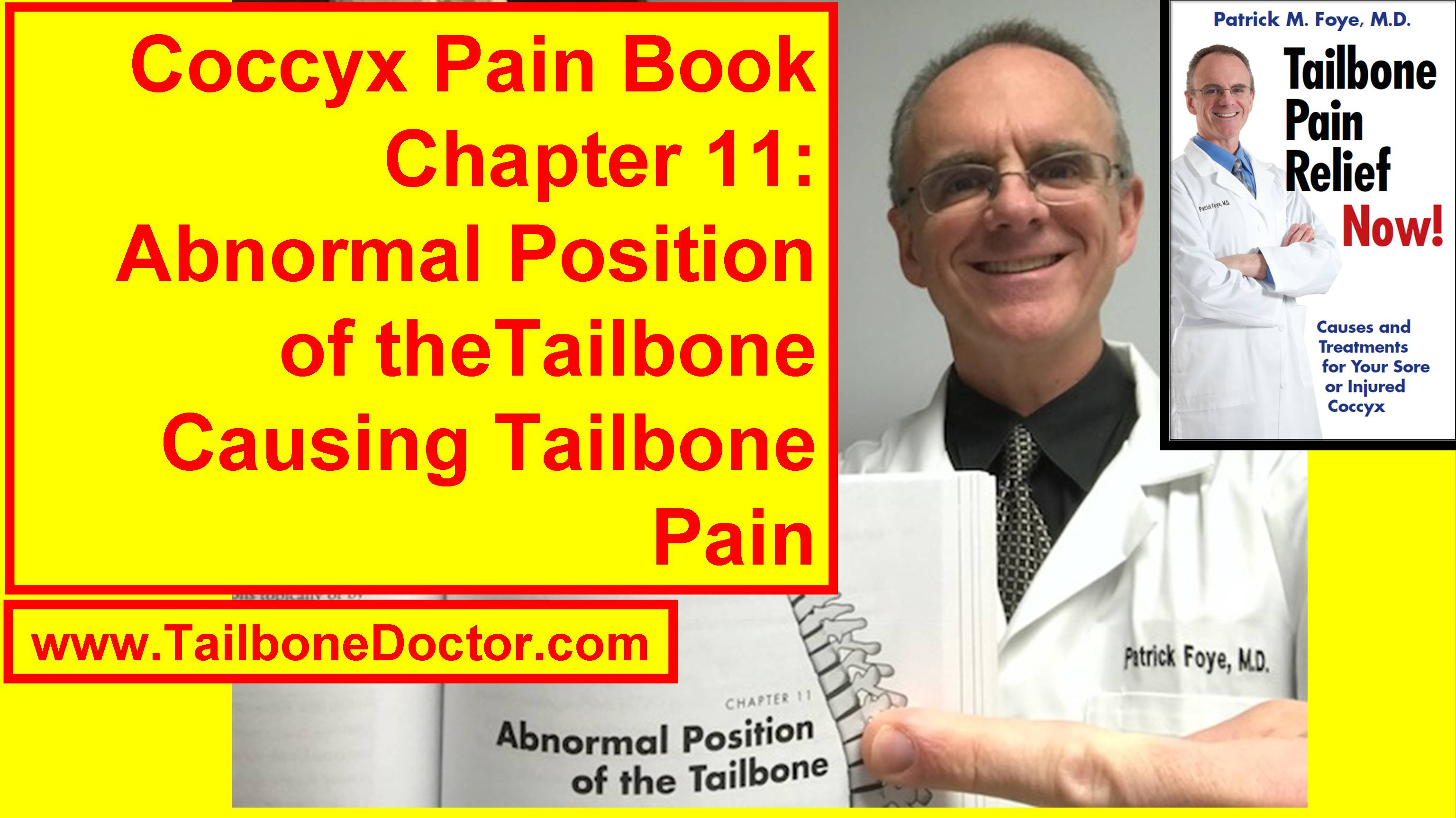
This is the next in a series of videos where I’m going chapter by chapter through my book “Tailbone Pain Relief Now!” providing the highlights of each of the chapters along the way.
And for this one we are at Chapter 11, which is “Abnormal Position of the Tailbone” or coccyx, as a cause of tailbone pain, coccyx pain or coccydynia.
And the general idea here is that there’s variability from person to person in terms of the shape and size and position of the tailbone.
And sometimes that can be something that causes tailbone pain, particularly when somebody is sitting down and putting their body weight onto the tailbone.
So in this video we’re just going to talk very briefly about a couple of the highlights from Chapter 11 of the book and specifically we are going to touch on two topics… Number one: what if the tailbone is too far forward? And number two: what if it is too far backwards?
So if the tailbone is flexed too far forwards, I am going to grab a model here and show you what may happen.
So here we have the lumbar spine up here and then down here is the sacrum and down here is the coccyx or tailbone.
And sometimes the tailbone can swing too far forward and if it does if it’s flexed too far forward, where my finger is now, for example, it has the potential to cause certain problems.
Number one, the tailbone is too far pushed into the pelvis and what that means is that if the tailbone is here, then when you have a bowel movement, the stool collects in the rectum which is right in this area.
So some people will have tailbone pain because the coccyx or tailbone is flexed too far forward, it’s actually blocking or obstructing the area where stool collects in the rectum to have a bowel movement.
And those people, as the stool collects, will start to have soreness and pain and aching.
And also, while you’re having a bowel movement it may be painful because the stool is pushing down through the rectum and is pushing that tailbone out of the way.
So that’s a tailbone that is flexed too far forwards.
Sometimes you can actually see that on the M.R.I. studies, there’s a particular view that needs to be done that’s unfortunately usually not done with a standard or typical pelvic MRI.
But if it’s specifically requested, there is a view that can be done that will show this nicely.
And it can show if the tailbone is sort of indenting into the rectum, that lowest part of the colon or the lowest part of the large intestine.
So that’s if a tailbone is flexed too far forward.
What if a tailbone is extended too far backwards?
This particular model does not let me really crank on it to show that, because I don’t want to break the model.
But if the tailbone instead of curving too far forward or being in its kind of normal slightly forward position, if the tailbone was instead extended backwards, well that causes a different set of problems.
Number one is that when you sit, the tailbone is sticking out.
And by sticking out… then when you go to sit… if this is the chair that you’re sitting on, that tailbone that is… (I can actually swing this model around I guess and show you… here would be if the tailbone was extending backwards).
Now instead of the tailbone being out of the way when you go to sit down on the chair, the tailbone now is basically contacting the chair.
And instead of the normal sort of rocker bottom aspect of the coccyx where it’s like the bottom of a rocking chair, instead if that lowest segment is tilting back, you can see that that is not going to be able to perform that rocking motion smoothly.
And instead it’s going to be pinching against the chair that you’re sitting on. So that’s going to pinch the skin in that area.
It can cause pain and inflammation there and it can also transmit those forces or pressures upstream. Just like if you ever caught a baseball or football or basketball and sort of jammed your finger while you’re catching it. You know that by doing that you kind of jam the fingers and then you can get pain within those joints.
Well similarly here if you’re sitting on a tailbone that’s extended backwards, you can sort of jam the joints there and have pain not just where the tailbone is contacting the chair but also at the joints up along the way.
So that’s a tailbone that’s extending too far backwards.
It’s important to have the imaging studies to see what is the position of the tailbone in any given patient who has tailbone pain. And that’s also going to help you to know what type of treatment might be appropriate and what types of problems those patients may run into.
I should also mention that for the patient who had the tailbone very far forward, that tailbone may be extending into the pelvis in a way that blocks or obstructs the birth canal.
So if the tailbone is hooked very far forward and it’s in the way of the birth canal that the child passes through or baby passes through during childbirth or labor and delivery then that can cause problems during childbirth both for the mom and potentially even for the baby.
So again it’s important to know the position of the tailbone in order to know what you should do about it and what the situation would be in those different scenarios.
This is showing a tailbone that is hooked and heading kind of far forward in that way.
So anyway, those are just a few highlights from Chapter 11 of the book.
If you’re looking for a full copy of the book, either the e-book which is the electronic book, or the paperback copy, it’s 272 pages all about the coccyx.
You can get that on Amazon.
But the easiest way perhaps is to just go to the website www.TailboneBoook.com and that will have the links directly to the Amazon pages that have this; depending on what country you’re in you click the appropriate link that matches there. That’s one way to find it.
You can also find me online or if you’re interested in coming for an evaluation here at the Coccyx Pain Center. The easiest way to do any of those things or to find other educational content and videos and articles that I have on tailbone pain is to find me at www.TailboneDoctor.com.
If you have comments or thoughts or questions about an abnormal position of the tailbone, the topic from this chapter of the book, post your comments down below.
I will be interested to read those and hopefully respond to those.
And others I am sure will be interested to read and respond to those as well.
So I look forward to your comments down below.
Okay, bye bye.
Here is the actual VIDEO:
Here is the screenshot thumbnail image for the video:
Chapter 11 of Tailbone Pain Book, Abnormal Coccyx Position Causing Coccyx Pain
To get your copy of the book “Tailbone Pain Relief Now!” go to: www.TailboneBook.com
For more information on coccyx pain, or to be evaluated at Dr. Foye’s Tailbone Pain Center in the United States, go to: www.TailboneDoctor.com
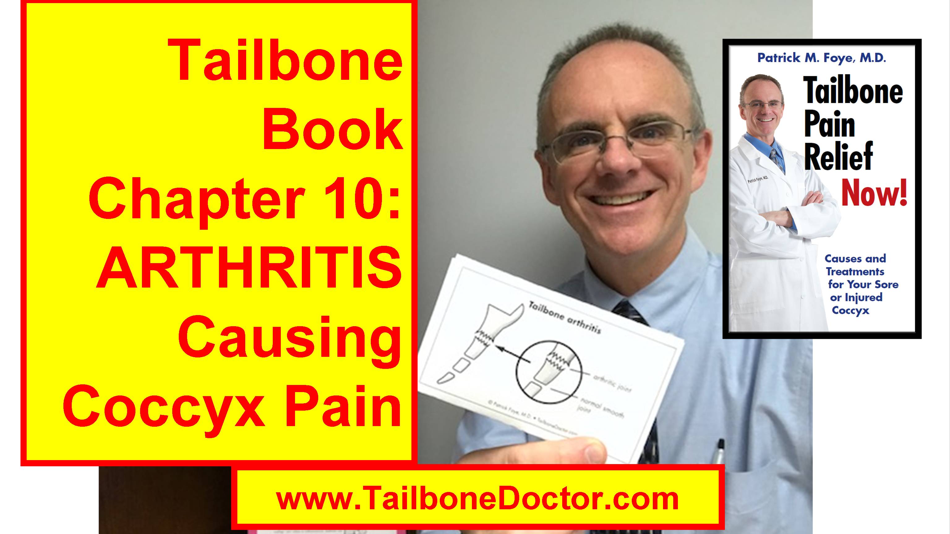
In this video we’re going to talk about ARTHRITIS as a cause of tailbone pain.
And this is the next in my series of videos going chapter by chapter through my book “Tailbone Pain Relief Now!”
I’m just doing short videos that give a glimpse as to some of the content that’s within each chapter.
And presenting it here in a video format so that it allows us to have an opportunity to interact.
And you can post your comments or questions or thoughts or your experiences with arthritis related to the tailbone down below.
And it gives us all a chance to interact down in the comments section.
So again back to the book.
We’re looking now at Chapter 10 within the book which is Arthritis of the Tailbone.
And most people are familiar with arthritis as a cause of pain in other parts of the body.
So you may have arthritis at your knee or at your hip.
Many people have arthritis within the small joints within their fingers.
And similarly people can have arthritis within the small joints within the coccyx.
And unfortunately many, many physicians forget to think about arthritis as a cause of coccyx pain.
Even though they would know enough to think about arthritis as a cause of pain in your fingers or your hip or your knee.
So let’s take a look for comparison again.
Just showing here where one joint meets the next… where one BONE, rather, meets the next.
That’s the joint.
And normally there’s a nice smooth appearance between one end of the one bone and the next.
But when there’s arthritic changes that happen that make it look abnormal.
And to show you a illustration from my book, this kind of overdramatizes the way the illustrator happened to draw it here.
But it makes the point which is you can see here the bones of the of the coccyx up one on top of the other.
And down in these lower joints in this example you can see that the bone surfaces are nice and smooth.
The bones may have a nice squared off appearance at the joint where one bone meets the next bone.
But at this bone up here you can see it doesn’t look so nice and neat and smooth anymore.
Instead it looks very kind of jagged or hazy or ragged around the edges.
And that’s because there’s arthritis at that joint.
There’s wear and tear at that joint.
Now this is sort of drawn in a very dramatic way for the illustration in the book.
It’s probably in most cases a little more subtle.
Something more like this where again over here you would have a nice neat joint as shown here as opposed to the arthritis.
Showing that the wear and tear within the joint over there on this part of the image.
So also at the edge of the joint you may have what’s called an osteophyte which is a little bit of a lip where the bone gets a little bit wider at that margin at the joint that has the arthritis.
So that’s a little bit about arthritis and some of the questions that come up about arthritis.
Well there are different types of arthritis.
The most common is osteoarthritis, which is also referred to as “degenerative joint disease.”
And this is normal wear and tear.
All of us go through life and we move around.
We put weight onto our joints.
Whether that’s by walking or running or sitting we’re putting weight onto our different joints throughout the body.
And some wear and tear happens at the joint over time.
And that wear and tear is referred to as arthritis.
The scenario can also happen where somebody can have trauma that causes arthritis.
Very commonly, you have a football player who has trauma to the knee and then at that knee joint they end up having arthritis much, much sooner… (many, many years or decades soone ) than they have normal aging arthritis at the same joint on the opposite side of the body.
So at the tailbone you can have trauma where maybe you had a slip and fall.
You landed on the tailbone.
You had an injury to the tailbone many, many, many years ago.
And it sort of did okay for a while.
Maybe for years or decades.
But arthritis starts to set in at that injured joint over time.
So that’s where you have trauma causing arthritis.
So whether the arthritis is caused by trauma or whether it’s just the micro-trauma of gradual wear and tear over the years, either way you can have arthritis at the tailbone.
The arthritis can cause pain because instead of having a nice smooth joint where one joint meets the next you have this irregular surface and that irregular surface is going to be associated with some extra friction now between one bone and the next and with any movement there.
And that can be quite painful for patients at the tailbone, just as it can be painful when they have arthritis in other parts of the body as well.
The other things we can talk about with arthritis is treatment of arthritis and diagnosis of arthritis.
So in terms of diagnosis really you need imaging studies to diagnose arthritis.
Whether that’s at the tailbone or at other parts of the body.
Most commonly this involves getting x-rays.
But you can get information from MRI and CT scans as well.
But really the x-rays are more or less the typical gold standard for diagnosing typical arthritis.
The x-rays need to be done in the proper way.
And what I mean by that is that it’s very, very common that the x-rays or other imaging studies at the tailbone will not be done in a way that adequately shows the tailbone.
So the imaging studies may be done of the lumbar spine which is higher up.
Or the imaging studies ARE done of the tailbone, BUT it doesn’t do the image at the proper angle in order to see the joints nice and clearly in order to make the diagnosis of arthritis.
So it becomes really important when the imaging studies are being done that the ordering physician understands and has experience in treating tailbone pain and orders the proper tests.
And also that the radiology technician does the tests properly.
And then that the radiologist that’s reading the images actually looks at the tailbone and comments on it and evaluates for some of these things.
And unfortunately it’s very, very common that there’s breakdown at each and every one of those steps along the way.
So although if I had arthritis within my finger the x-rays would be done properly.
The radiologist would read it properly etc.
But if I had arthritis instead of at my finger, if I had the arthritis at my tailbone, then often the system just sort of breaks down because most people are not familiar with treating tailbone pain.
As far as treatment of arthritis, the first step in treatment is actually having an accurate diagnosis in the first place.
And that’s why the imaging studies and the evaluation are so important.
The treatment sometimes can involve putting medication locally at the spot such as by a small local injection.
But we’re going to get into treatment in another chapter later within the book.
So we’ll have a chance to talk more about that there.
But for this purpose let’s just say that unless you make an accurate diagnosis in the first place, it’s very difficult to have a logical treatment plan.
Because if you just are labeled as having “tailbone pain” overall, which they call coccydynia, withOUT having a diagnosis made as to what’s CAUSING the tailbone pain, well then you don’t have a clear direction for what way you’re going to go with your treatment options.
Or you may have an injection done but they don’t do the injection at the specific joint within the coccyx that has the arthritis because they haven’t made that diagnosis in the first place.
So I hope that information is helpful about arthritis in the tailbone.
Definitely post your comments, questions, and stories related to tailbone arthritis down below.
So that gives us a chance to read and comment back on those.
If you’re looking for a copy of the entire book for more information on arthritis of the tailbone and lots of other things about the tailbone, the book is available online.
The easiest way to get it online is to go to the website www.TailboneBook.com and that’ll have the links to different amazon pages for different countries etc.
You can get it as a printed book which is 272 pages in paperback, or you can get the electronic book, the eBook version, if that’s easier for you.
You can get that pretty much anywhere in the world that has internet access.
You can download the electronic book and have access to all of the same information.
If you’re looking specifically for me online, the easiest place to find me is at the website www.TailboneDoctor.com.
So if you’re looking for more educational content, I have lots of educational videos and online articles and information written specifically about the tailbone and tailbone pain.
You can find all of that there.
Or if you’re looking to come to the Tailbone Pain Center for evaluation and treatment, all of that information is there as well at www.TailboneDoctor.com.
So I hope the information is helpful for you.
I’ll be looking for your comments down below regarding arthritis at the tailbone.
Bye bye.
Here is the actual VIDEO:
Here is the screenshot thumbnail image for the video: