Someone recently asked me whether it was possible for someone to have coccyx pain being caused by TWO different causes in the same patient.
Yes, it is certainly possible for patients to have more than one location of coccyx problem.
For example, I frequently see patients who have both the most common cause of coccyx pain (hypermobility, diagnosed by sitting-versus-standing x-rays) and they may ALSO have the 2nd most common cause of coccyx pain, which is a bone spur at the distal/lowest tip of the coccyx.
Or, some patients may have pain being caused by hypermobility or other abnormalities at TWO different joints within the coccyx, not just one joint.
Or, a patient may have arthritis at the upper coccyx, and tables for at the lower coccyx.
Other combinations are also possible.
The best way to help figure all this out is to have a thorough and thoughtful medical evaluation by a physician who is experienced at evaluating and treating coccydynia (coccyx pain, tailbone pain).
GET THE BOOK: To get your copy of the book “Tailbone Pain Relief Now!” go to: www.TailboneBook.com or go to Amazon
COME FOR RELIEF: For more information on coccyx pain, or to be evaluated in-person at Dr. Foye’s Tailbone Pain Center in the United States, go to: www.TailboneDoctor.com
– Patrick Foye, M.D., Director of the Coccyx Pain Center, New Jersey, United States.
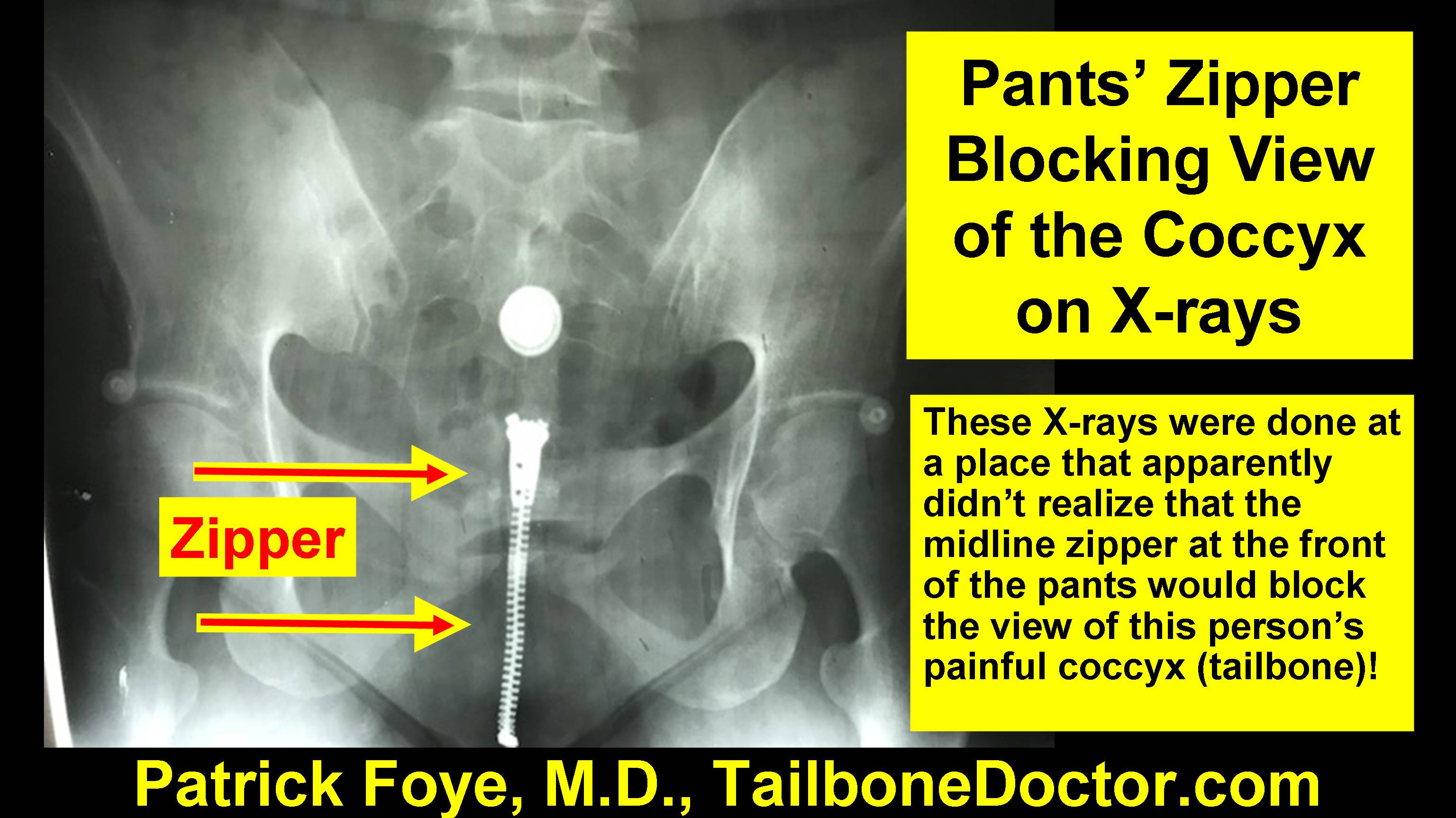
Pants’ Zippers Blocking View of the Coccyx on Tailbone X-rays
Many radiology imaging centers have no idea how to properly perform x-rays of the coccyx for people who have tailbone pain (also called coccyx pain or coccydynia).
Sometimes even the most basic problems occur.
I have seen many, many patients over the years where the prior x-rays were done in such a way that the tailbone could not be seen at all.
Sometimes the imaging studies do not include the coccyx because the images are of the wrong body region.
Sometimes, like in this example, the images are done of the correct body region, but the patient is wearing denim jeans or other pants with a zipper that completely blocks the view of the coccyx on the frontal view (also called the A-P, or anterior-posterior, view).
This could have been easily avoided by having the patient change into a gown, or shorts without a zipper, before the x-rays were taken.
Instead, these patients undergo poorly done testing.
Not only does this waste time and money by doing a test that gives no useful information. Even worse is that often the treating doctor fails to look at the images themselves, so they don’t even realize there was a problem. Then, the patient is just told that their coccyx x-rays did not reveal any coccyx abnormalities. But, in fact, the coccyx x-rays failed to even adequately show the coccyx!
This may sound crazy that these things would happen. But patients traveling to see me from all around the country and it is unfortunately VERY common that the previous imaging studies have these kinds of problems. I discover these kinds of problems in my patients’ prior imaging studies essentially everyday.
Here is the video:
Here is the screenshot image from the video:
Pants Zipper Blocks View of the Coccyx, Tailbone, on X-rays
To get your copy of the book “Tailbone Pain Relief Now!” go to: www.TailboneBook.com
For more information on coccyx pain, or to be evaluated at Dr. Foye’s Tailbone Pain Center in the United States, go to: www.TailboneDoctor.com
Text transcribed from the video above:
This video is about zippers on your pants that can be blocking the view of the coccyx on x-rays.
So for people with tailbone pain, if they have an x-ray done of the coccyx, they can have zippers on their pants that can get in the way of the x-rays (which could happen if they are not changed into a gown to have the x-rays taken).
I’m Dr. Patrick Foye.
I’m the Director of the Coccyx Pain Center here in the United States.
And I’ve been treating people with tailbone pain for over 20 years.
I’ve treated thousands of patients.
And unfortunately from time to time there will be somebody who comes in and they bring their x-rays with them that they had done previously at an outside institution… and we’ll take a look at the x-rays, take a look to see if they show the coccyx or tailbone.
And what happens is what happened here, which is the tailbone is hidden behind the zipper of the patient’s pants.
So, this is a view of the pelvis right here.
So I don’t know if this will show up, I’m kind of in a dark room at the moment, but I’m holding up an anatomic model of the pelvis and at the back of the pelvis you can see the sacrum and coccyx there.
But on this x-ray, here’s our pelvis, the sacrum is back here, the coccyx would be hidden right behind the zipper.
It’s also in this case hidden behind the pubic bone but for purposes of this x-ray and the point of this video, even if the image was appropriately tilted to show the coccyx the zipper would still be blocking the view of the coccyx.
And it’s really a shame because people have tailbone pain and they suffer for months and years often without an accurate diagnosis.
And one of the reasons why they suffer for years without an accurate diagnosis is because so many places do the wrong imaging studies or do the imaging studies the wrong way.
So in this patient again the exact area that the study was being done for (which was tailbone pain) the tailbone is hidden behind the zipper of the person’s pants.
And here’s the buckle or the button or rivet or whatever just above the zipper at the front of the person’s pants.
And you can see some of the other rivets here and here from the side pockets of the pants.
So again this could be very easily fixed by just having a patient change into a gown, or into a pair of shorts even that does not have any buttons or zippers in the patient’s clothing.
And especially that if it did have buttons or zippers that you do the imaging studies in such a way that it’s not blocking the area that you are most interested in for that patient.
So that’s the general take-home message.
The other thing that can be done of course is to do the x-ray from the side.
So that instead of only taking the x-ray from that front view (where you can see right now the pubic bone is blocking the view now the tailbone comes in but if the zipper was there it would still be blocked) but the other thing you can do is to take the image from the side which is more aligned kind of like this which is called a lateral view in medical lingo.
So those are a couple of points about the x-rays of the coccyx and avoiding having a zipper get in the way of the coccyx or tailbone on your x-rays.
For more information, you can find me online at www.TailboneDoctor.com or on Twitter @TailboneDoctor.
And if you have questions about this, you can post them down below this video.
COME FOR RELIEF: For more information on coccyx pain, or to be evaluated in-person at Dr. Foye’s Tailbone Pain Center in the United States, go to: www.TailboneDoctor.com
– Patrick Foye, M.D., Director of the Coccyx Pain Center, New Jersey, United States.
e-Book on Coccyx Pain, Tailbone Pain, for Physicians, from the First International Coccyx Pain Symposium in 2016
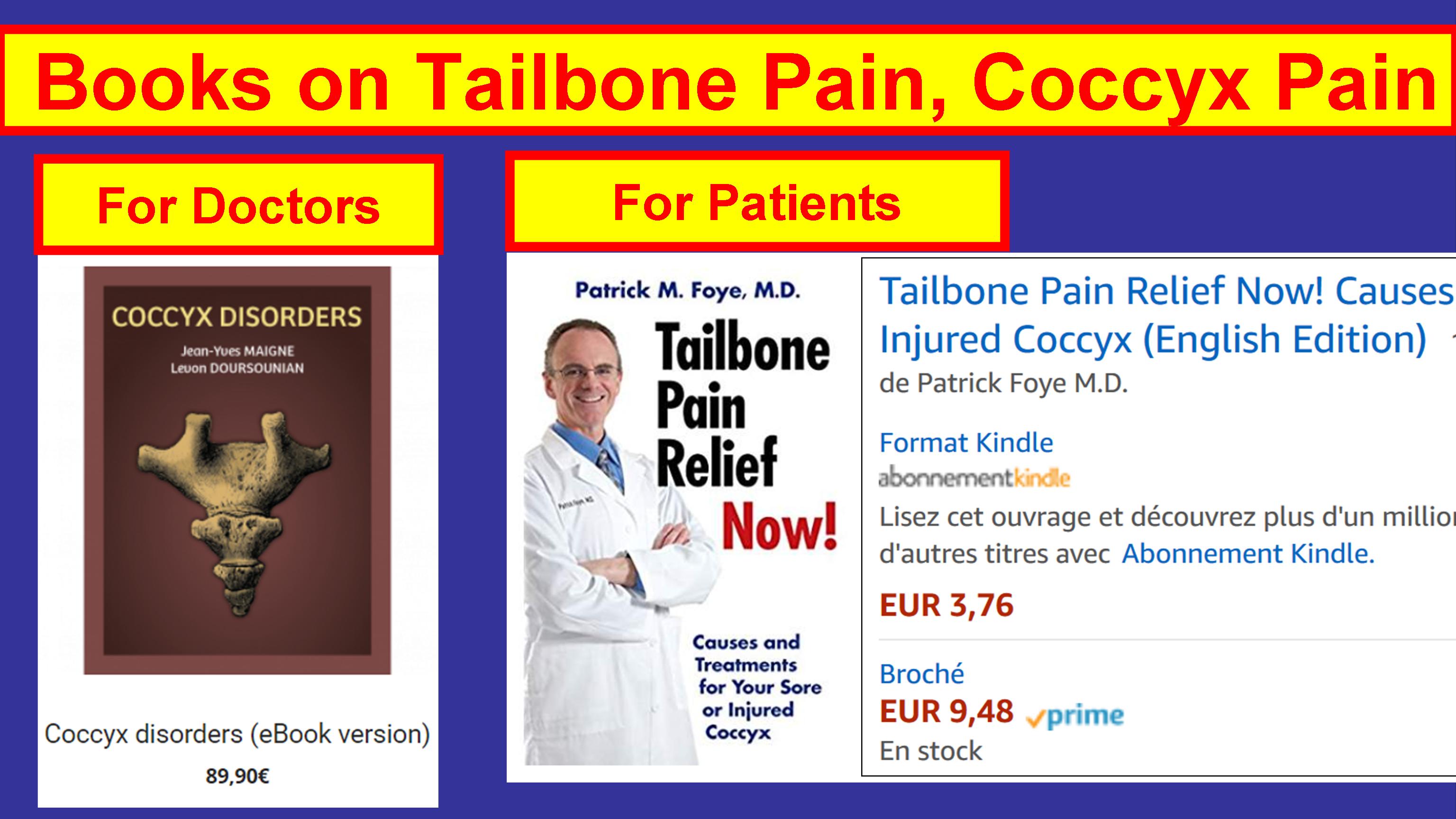
Coccyx Pain Books, For Doctors versus For Patients
Dr. Foye was Interviewed by Dr. Elif Gürkan, from Istanbul, Turkey, regarding Coccyx Pain (coccydynia, tailbone pain, koksidini, koksiks ağrı).
They discussed coccydynia (koksidini, coccyx pain, tailbone pain, koksiks ağrı).
If you have tailbone pain and live in Turkey… You can find Dr. Gürkan’s website online at: www.elifgurkan.com
Here is the video, which is in both English and Turkish:
Screen capture from the video:
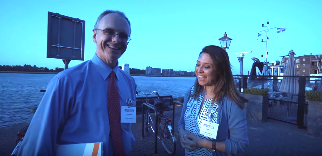
Patrick Foye, MD, was interviewed in Holland, the Netherlands, in June 2018, by Dr Elif Gürkan, from Istanbul, Turkey
GET THE BOOK: To get your copy of the book “Tailbone Pain Relief Now!” go to: www.TailboneBook.com or go to Amazon
COME FOR RELIEF: For more information on coccyx pain, or to be evaluated in-person at Dr. Foye’s Tailbone Pain Center in the United States, go to: www.TailboneDoctor.com
– Patrick Foye, M.D., Director of the Coccyx Pain Center, New Jersey, United States.
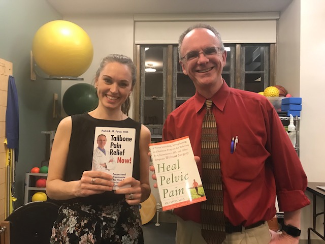
Pelvic Floor Physical Therapy can be immensely valuable for many patients with certain types of pelvic pain.
I was reflecting this weekend and feeling grateful for having met and collaborated with so many wonderful Pelvic Floor Physical Therapists over the years.
It has been wonderful sharing patients in common, or giving lectures to the therapists, or attending lectures that they have given. They have taught me a great deal.
I’m continuing to learn more and more about the important role that Pelvic Floor Physical Therapy can play in the treatment of various types of pelvic floor dysfunctions.
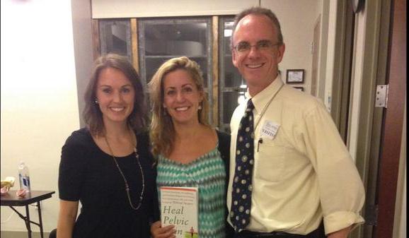
A few of the Pelvic Floor Physical Therapists are shown below.
At Beyond Basics Pelvic Floor Physical Therapy, NYC, Stephanie Stamas, Amy Stein, and Patrick Foye MD. October 2014
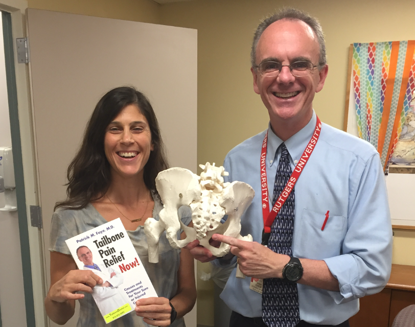
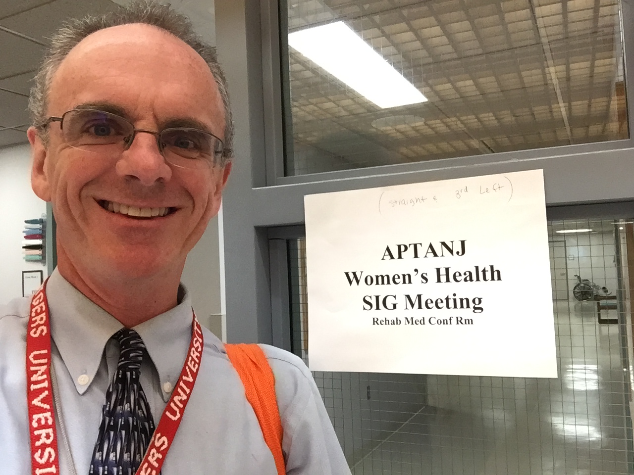
Pelvic Floor Physical Therapists with Patrick Foye MD, after his lecture on Tailbone Pain, for the American Physical Therapy Association’s New Jersey Chapter, Women’s Health Special Interest Group. 2016.
Niva Herzig PT with Patrick Foye MD, after Dr. Foye’s lecture on Tailbone Pain, for the American Physical Therapy Association’s New Jersey Chapter, Women’s Health Special Interest Group. 2016.
Patrick Foye MD, gave a lecture on Tailbone Pain, for the American Physical Therapy Association’s New Jersey Chapter, Women’s Health Special Interest Group (Pelvic Floor Physical Therapists). 2016.
Pelvic Floor Physical Therapists’ Coccyx Conference. Herman and Wallace. In Florida, March 2017.
Dr Foye Gives a Lecture on Coccyx Pain (Tailbone Pain) to Pelvic Floor Physical Therapists. In Florida, March 2017.
Patrick Foye MD with Netherlands Pelvic Floor Physical Therapist Marijke Slieker. At the International Coccyx Pain Symposium, in Holland, 2018.
Rivki Chudnoff (from Hamakom PT), along with Stacey Futterman Tauriello & Kara (both from @5pointPT),. In Newark, New Jersey, July 2018.
Stephanie Stamas PT DPT, with Patrick Foye MD, after her Pelvic Pain lecture, NYC, March 2019.
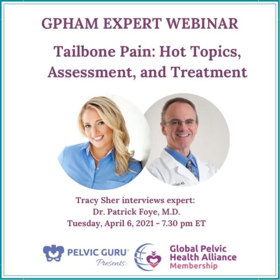
Tailbone Pain Lecture by Patrick Foye, MD, at ProTouch PT, Aug 2019, Cranford NJCoccyx Pain, Tailbone Pain, Webinar by Patrick Foye MD, for Pelvic Guru, GPHA, April 2021
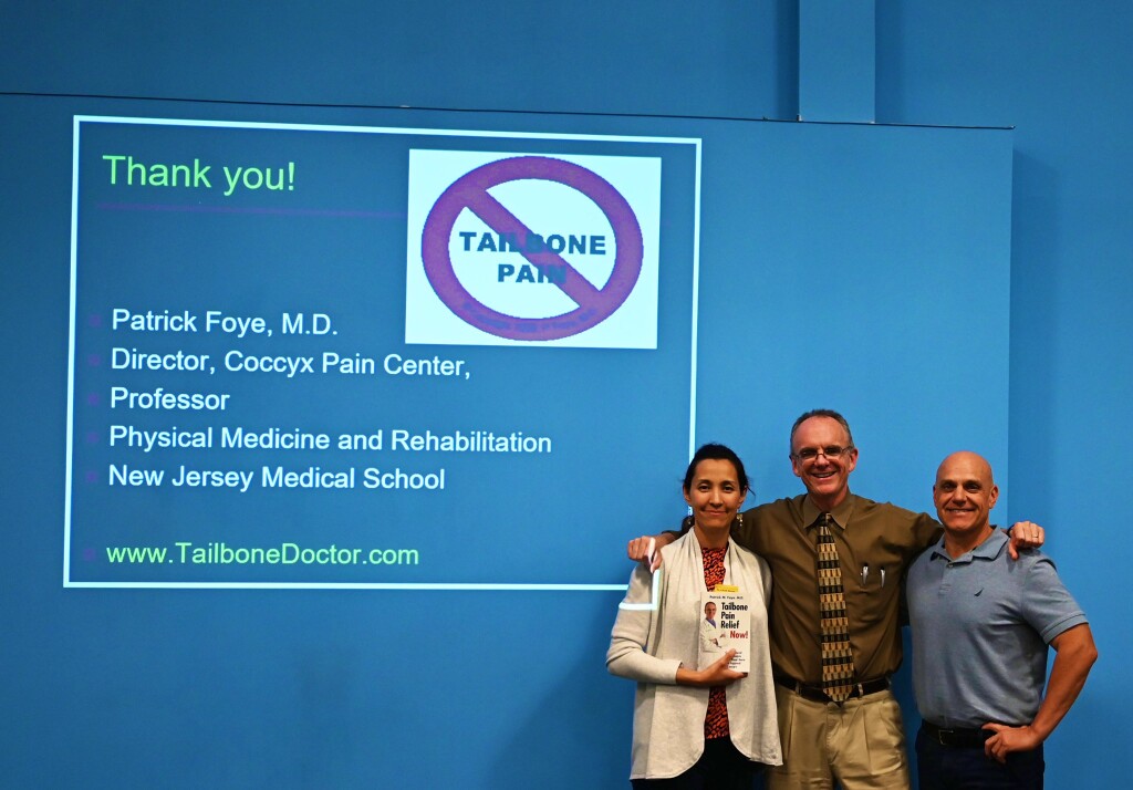
Looking forward to learning more and collaborating more over the coming years, as we all strive together to relieve pain and suffering for our patients.
– Patrick Foye, M.D., Director of the Coccyx Pain Center, Rutgers New Jersey Medical School. www.TailboneDoctor.com
“STIR” images and “T2” images on MRI are essentially like filter settings where fluid shows up bright.
Some amount of fluid is normal, since many of our body tissues contain significant fluid content (for example, our fat cells [adipose] have significant fluid content).
Also, we of course have the fluid we call urine within our urinary bladder, and cerebrospinal fluid within our spinal canal, and fluid within our bowels.
But sometimes the STIR images or T2 images show an *increased* or *abnormal* amount of fluid, or a fluid signal in a place where it is not normally seen.
Commonly this extra fluid signal is due to inflammation.
Less commonly it can be things like infection (but that would usually be apparent clinically, meaning that someone would often have other symptoms like fever, redness/warmth of the skin overlying the area, etc.).
A fluid-filled cyst (such as a pilonidal cyst) would often also show up bright on T2 and STIR images.
So, where there is increased signal on T2 and STIR images, we often try to figure out whether this is caused by inflammation or if it is caused by something else.
Overall, the cause is frequently inflammation.
If we believe that the increased signal was due to inflammation, then the next step is to try to figure out what is causing the inflammation.
For example, sometimes there will be inflammation at/around a joint that is unstable (coccygeal dynamic instability).
When there is inflammation around the lowest coccygeal bone, often that can be associated with an underlying bone spur at that lowest tip of the coccyx. But, often the bone spur will be missed if the MRI was not done in a way that included sagittal images in the midline (basically sagittal is an image of what it would look like if we were to slice and someone right down the middle into a left half and the right half). Also, the sagittal images to look for a bone spur would typically need to be done with the “T-1” filter setting, since that is the best MRI filter setting to show bony structures.
All of this assumes that the radiologist or treating physician took the time to carefully look at the MRI images and to specifically look for these kinds of abnormalities at the coccyx.
This is especially important in patients with coccyx pain (coccydynia, coccyx pain).
Here at the Coccyx Pain Center, I see hundreds of new patients per year where their prior imaging studies either failed to include the coccyx at all, or fail to include the correct types of slices, or fail to include the correct filter settings, or where the radiologist and treating physician failed to closely look for these kinds of findings.
People suffering from coccyx pain should educate themselves about these issues, so that they can better advocate for themselves in getting the best medical care possible, even if their local treating physicians may not be particularly familiar with treating coccydynia.
GET THE BOOK: To get your copy of the book “Tailbone Pain Relief Now!” go to: www.TailboneBook.com or go to Amazon
COME FOR RELIEF: For more information on coccyx pain, or to be evaluated in-person at Dr. Foye’s Tailbone Pain Center in the United States, go to: www.TailboneDoctor.com
– Patrick Foye, M.D., Director of the Coccyx Pain Center, New Jersey, United States.
I like the way this video is really nicely illustrated.
It is just showing the basics, without much detail, but that is a perfect starting point for patients who are just first learning about their pelvic floor.